Neuroscience

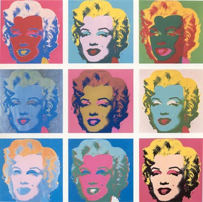
Move over, Marilyn Monroe neurons and Halle Berry neurons... The cellular media darlings of action observation and action execution would like to join you in the human hippocampus and surrounding medial temporal lobe (MTL) areas critical for memory.
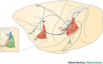
"What?" you say. "Direct evidence for the existence of mirror neurons has been obtained from single cell recordings in monkeys in specific brain regions. These include the ventral premotor cortex (area F5) and the inferior parietal lobule (Rizzolatti & Sinigaglia, 2010), not the hippocampus!"

Figure 1 (Rizzolatti & Sinigaglia, 2010): The parieto-frontal mirror network. Lateral view of the macaque brain. The coloured areas represent the areas of the parieto-frontal circuit containing mirror neurons: the ventral premotor cortex (area F5), area PFG (located between parietal areas PF and PG) and the anterior intraparietal area (AIP)... The parieto-frontal circuit receives high-order visual information from areas located inside the superior temporal sulcus (STS) and the inferior temporal lobe (IT). Neither of these temporal regions has motor properties. The parieto-frontal circuit is under control of the frontal lobe (area F6 or pre-supplementary motor area and the ventral prefrontal cortex (VPF)). The inset provides an enlarged view of area F5. IAS, inferior limb of the arcuate sulcus; LIP, lateral intraparietal area; VIP, ventral intraparietal area.
Yet we've been led to believe that a new study published in Current Biology (Mukamel et al., 2010) has recorded from mirror neurons in the human brain for the first time. Last Friday, BPS Research Digest asked:
The experimental protocol consisted of 3 tasks: Grasp, Facial expressions and Control.
 In each brain region, only a minority of cells responded with an increased (or decreased) firing rate during both observation and execution of the same action. Percentages of these Observation/Execution neurons ranged from a low of 2% in dorsal anterior cingulate cortex to 11% in hippocampus, 12% in parahippocampal gyrus, and 14% in supplementary motor area. The MTL regions also contain neurons that respond during the spontaneous recall of episodic memories (Gelbard-Sagiv et al., 2008). So how can you tell if a neuronal response in the current experiment is related to memory recall or to action observation/execution? You can't, but that doesn't matter!
Good point.
Another issue is whether a given single- or multi-unit recording responded by increasing or decreasing its firing rate relative to the passive condition. It could be either, or both:
Also notable is that the paper did not refer to previous results from the same lab on Marilyn Monroe neurons (Quian Quiroga et al., 2009) and Halle Berry neurons (Quian Quiroga et al., 2005).1 The new "mirroring cells" are apparently intermixed with individual neurons that show hyperspecific responses to pictures of celebrities (taken from various angles, in and out of character) and even to their printed names and voices. In each brain region, about 10% of the cells were responsive to stimulation of any sort. Of these minority neurons, 0% in parahippocampal cortex, 14% in amygdala, 35% in entorhinal cortex, and 38% in hippocampus showed "triple invariance" to presentation as pictures, sound, and text (Quian Quiroga et al., 2009).
The hyperspecific neuronal responses included a Jennifer Aniston+Brad Pitt cell (not Aniston alone), a Pamela Anderson cell that responded to a caricature of her and to her printed name, and a Kobe Bryant cell. A specific double dissociation was reported between a Halle Berry neuron and a Mother Teresa neuron (i.e, one cell showed a response to Halle Berry but not Mother Teresa, and the other cell showed a response to Mother Teresa but not Halle Berry).
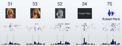
Below is one of my favorites, the rare Robert Plant neuron that responds to images, sounds, and text depicting the former Led Zeppelin singer. I wonder what would happen if he had a closed shirt and shorn hair in some of the pictures?

Figure S17 (Quian Quiroga et al., 2009). A single unit in the entorhinal cortex selectively activated by pictures, sound and text presentations of Robert Plant, singer of the band ‘Led Zeppelin’, which was known to the patient.
But what about hybrid Halle Berry mirror neurons? How does one integrate the results from all these studies? Who's to say that you couldn't find a Jennifer Aniston action observation/action imitation neuron if you looked hard enough? Would it still be considered a "mirroring neuron" if Lisa Kudrow did not elicit the same response? What if all the cast members of Friends could evoke the observation/imitation response, but not the cast members of Seinfeld?
In conclusion, Mukamel et al., (2010) have this to say about their "mirror neuron" results:
Footnote
1 One of the Fried lab papers was cited in passing but only to say how the new "mirror neuron" results were not due to representational invariance of grasping or smiling, because cell firing rates did not change in the passive control condition. However, the lack of a significant response in the control condition was required before including a cell(s) in the action execution bin. I can imagine that quite a few neurons responded in a condition where subjects were told, "covertly read the words and refrain from making hand movements or facial gestures" -- especially in the SMA and pre-SMA, which are involved not only in motor planning but in motor inhibition as well.
References
Gelbard-Sagiv H, Mukamel R, Harel M, Malach R, Fried I. (2008). Internally generated reactivation of single neurons in human hippocampus during free recall. Science 322:96-101.
Mukamel, R., Ekstrom, A., Kaplan, J., Iacoboni, M., & Fried, I. (2010). Single-Neuron Responses in Humans during Execution and Observation of Actions. Current Biology DOI: 10.1016/j.cub.2010.02.045
Quian Quiroga, R., Kraskov, A., Koch, C., & Fried, I. (2009). Explicit Encoding of Multimodal Percepts by Single Neurons in the Human Brain. Current Biology, 19 (15), 1308-1313 DOI: 10.1016/j.cub.2009.06.060
Quiroga, R., Reddy, L., Kreiman, G., Koch, C., & Fried, I. (2005). Invariant visual representation by single neurons in the human brain. Nature, 435 (7045), 1102-1107 DOI: 10.1038/nature03687
Rizzolatti G, Sinigaglia C. (2010). The functional role of the parieto-frontal mirror circuit: interpretations and misinterpretations. Nat Rev Neurosci. 11:264-74.

- Mirror Neuron Death March
Above image: Jim Peters almost wins the marathon Vancouver, 7 August 1954, with mirror neurons by Rizzolatti & Craighero (2004). Greg Hickok at Talking Brains has a series of posts dismantling the mirror neuron theory of action understanding. Actually,...
- Mirror Neurons In Primary Motor Cortex?
The mirror neurons, it would seem, dissolve the barrier between self and others. I call them "empathy neurons" or "Dalai Llama neurons". -- MIRROR NEURONS AND THE BRAIN IN THE VAT by V.S. Ramachandran Everyone knows what mirror neurons are,...
- Spindle Neurons: The Next New Thing?
In a neuroanatomical tour de force, Nimchinsky and colleagues (1999) obtained access to samples of the anterior cingulate cortex (and other cortical regions) from 28 different primate species, from prosimians to anthropoids to great apes to humans. They...
- Neuropsychology Abstract Of The Day: Mirror Neurons
Brain regions with mirror properties: A meta-analysis of 125 human fMRI studies Neuroscience Biobehavioral Rev. 2011 Jul 18. [Epub ahead of print] Molenberghs P, Cunnington R, Mattingley JB. Abstract Mirror neurons in macaque area F5 fire when an animal...
- Specialized Neurons Of The Medial Temporal Lobe
This study has been reported in the media in a number of different locations. Today, The New York Times includes a small report about it:A Neuron With Halle Berry's Name on It By SANDRA BLAKESLEE The New York Times Published: July 5, 2005 Scientists...
Neuroscience
Mirror Neurons Join Marilyn Monroe Neurons and Halle Berry Neurons in the Human Hippocampus

Move over, Marilyn Monroe neurons and Halle Berry neurons... The cellular media darlings of action observation and action execution would like to join you in the human hippocampus and surrounding medial temporal lobe (MTL) areas critical for memory.
"What?" you say. "Direct evidence for the existence of mirror neurons has been obtained from single cell recordings in monkeys in specific brain regions. These include the ventral premotor cortex (area F5) and the inferior parietal lobule (Rizzolatti & Sinigaglia, 2010), not the hippocampus!"

Figure 1 (Rizzolatti & Sinigaglia, 2010): The parieto-frontal mirror network. Lateral view of the macaque brain. The coloured areas represent the areas of the parieto-frontal circuit containing mirror neurons: the ventral premotor cortex (area F5), area PFG (located between parietal areas PF and PG) and the anterior intraparietal area (AIP)... The parieto-frontal circuit receives high-order visual information from areas located inside the superior temporal sulcus (STS) and the inferior temporal lobe (IT). Neither of these temporal regions has motor properties. The parieto-frontal circuit is under control of the frontal lobe (area F6 or pre-supplementary motor area and the ventral prefrontal cortex (VPF)). The inset provides an enlarged view of area F5. IAS, inferior limb of the arcuate sulcus; LIP, lateral intraparietal area; VIP, ventral intraparietal area.
Yet we've been led to believe that a new study published in Current Biology (Mukamel et al., 2010) has recorded from mirror neurons in the human brain for the first time. Last Friday, BPS Research Digest asked:
Is this the first ever direct evidence for human mirror neurons?The short answer to this question is "no" (unless you want to dilute the meaning of "mirror neurons" beyond recognition). To briefly summarize, the participants in the study were 21 patients with pharmacologically intractable epilepsy. Depth electrodes were implanted into their brains to monitor for seizures, in advance of a possible surgical intervention to remove the epileptic focus. The electrode locations were constrained by clinical considerations and included regions in the medial frontal cortex (supplementary motor area, anterior cingulate cortex) and the medial temporal lobe (amygdala, hippocampus, parahippocampal gyrus, entorhinal cortex). The ventral premotor cortex and inferior parietal lobe were not targeted.
...Although recordings from single cells in the brains of monkeys have identified 'mirror' neurons that respond both to the execution of a movement and the observation of another agent performing that same movement, the existence of such cells in humans has, up until now, been inferred only from indirect evidence, particularly brain imaging. Now Roy Mukamel and colleagues have provided what appears to be the first ever direct evidence, using implanted electrode recordings of single cells, for the existence of mirror neurons in humans.
The experimental protocol consisted of 3 tasks: Grasp, Facial expressions and Control.
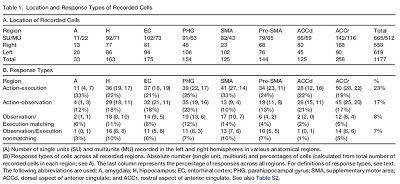
During Grasp, subjects were presented with video clips of a hand grasping a mug and with the words ‘Finger’ or ‘Hand’. They were instructed to grasp a mug with precision grip or whole hand prehension when the words ‘Finger’ or ‘Hand’, respectively, were presented and to simply observe when the video clips were played. During Facial expressions, subjects were presented with a picture of a smiling or a frowning face and with the word ‘Smile’ or ‘Frown’. They were instructed to perform the corresponding action when the words were presented and to simply observe when the pictures were presented... In the Control task, subjects were presented with the words used as cues in the Grasp and Facial expression parts of the experiment and were instructed to covertly read the words and refrain from making hand movements or facial gestures.Results are depicted in Table 1 below (click on image for a larger view).
 In each brain region, only a minority of cells responded with an increased (or decreased) firing rate during both observation and execution of the same action. Percentages of these Observation/Execution neurons ranged from a low of 2% in dorsal anterior cingulate cortex to 11% in hippocampus, 12% in parahippocampal gyrus, and 14% in supplementary motor area. The MTL regions also contain neurons that respond during the spontaneous recall of episodic memories (Gelbard-Sagiv et al., 2008). So how can you tell if a neuronal response in the current experiment is related to memory recall or to action observation/execution? You can't, but that doesn't matter!
The action observation/execution matching neurons in the medial temporal lobe may match the sight of actions of others with the memory of those same actions performed by the observer. Thus during action-execution, a memory of the executed action is formed, and during action-observation this memory trace is reactivated. This interpretation is in line with the hypothesis of multiple mirroring mechanisms in the primate brain, a hypothesis that can easily account for the presence of mirroring cells in many cortical areas."Now wait a minute," said Professor Patricia Churchland [as paraphrased by Prof. Greg Hickok in Talking Brains]. "If mirror neurons are all over the brain then don't they lose their explanatory power?"
Good point.
Another issue is whether a given single- or multi-unit recording responded by increasing or decreasing its firing rate relative to the passive condition. It could be either, or both:
Among the 68 action observation/execution matching cells [out of 1177 total cells recorded], 33 increased their firing rate during both observation and execution of a particular action. In contrast, 21 other neurons decreased their firing rate during both conditions. These types of responses have been previously reported in monkeys and birds. Furthermore, 14 neurons increased their firing rate during one condition and decreased it during the other.Really? Neurons can show inhibitory responses to observation and execution, or mismatched responses, and still be considered "mirror neurons"?
Also notable is that the paper did not refer to previous results from the same lab on Marilyn Monroe neurons (Quian Quiroga et al., 2009) and Halle Berry neurons (Quian Quiroga et al., 2005).1 The new "mirroring cells" are apparently intermixed with individual neurons that show hyperspecific responses to pictures of celebrities (taken from various angles, in and out of character) and even to their printed names and voices. In each brain region, about 10% of the cells were responsive to stimulation of any sort. Of these minority neurons, 0% in parahippocampal cortex, 14% in amygdala, 35% in entorhinal cortex, and 38% in hippocampus showed "triple invariance" to presentation as pictures, sound, and text (Quian Quiroga et al., 2009).
The hyperspecific neuronal responses included a Jennifer Aniston+Brad Pitt cell (not Aniston alone), a Pamela Anderson cell that responded to a caricature of her and to her printed name, and a Kobe Bryant cell. A specific double dissociation was reported between a Halle Berry neuron and a Mother Teresa neuron (i.e, one cell showed a response to Halle Berry but not Mother Teresa, and the other cell showed a response to Mother Teresa but not Halle Berry).
Below is one of my favorites, the rare Robert Plant neuron that responds to images, sounds, and text depicting the former Led Zeppelin singer. I wonder what would happen if he had a closed shirt and shorn hair in some of the pictures?

Figure S17 (Quian Quiroga et al., 2009). A single unit in the entorhinal cortex selectively activated by pictures, sound and text presentations of Robert Plant, singer of the band ‘Led Zeppelin’, which was known to the patient.
But what about hybrid Halle Berry mirror neurons? How does one integrate the results from all these studies? Who's to say that you couldn't find a Jennifer Aniston action observation/action imitation neuron if you looked hard enough? Would it still be considered a "mirroring neuron" if Lisa Kudrow did not elicit the same response? What if all the cast members of Friends could evoke the observation/imitation response, but not the cast members of Seinfeld?
In conclusion, Mukamel et al., (2010) have this to say about their "mirror neuron" results:
The functional significance of the mirror mechanism most likely varies according to the location of mirror neurons in different brain areas. For example, the mirror mechanism in the insula might underlie the capacity to understand a specific emotion (disgust) in others, whereas the mirror mechanism in the parietofrontal circuit may help understanding the goal of observed motor acts and the intentions behind them. Here we show cellular mirroring mechanisms in areas relevant to movement initiation and sequencing (SMA) and to memory (medial temporal lobe). Whereas these hypotheses have yet to be tested more carefully, these results demonstrate the presence of mirror mechanisms in humans at the single neuron level and in areas functionally different from the ones previously described in the literature.So are mirror neurons everywhere? Have they lost their explanatory power?
Footnote
1 One of the Fried lab papers was cited in passing but only to say how the new "mirror neuron" results were not due to representational invariance of grasping or smiling, because cell firing rates did not change in the passive control condition. However, the lack of a significant response in the control condition was required before including a cell(s) in the action execution bin. I can imagine that quite a few neurons responded in a condition where subjects were told, "covertly read the words and refrain from making hand movements or facial gestures" -- especially in the SMA and pre-SMA, which are involved not only in motor planning but in motor inhibition as well.
References
Gelbard-Sagiv H, Mukamel R, Harel M, Malach R, Fried I. (2008). Internally generated reactivation of single neurons in human hippocampus during free recall. Science 322:96-101.
Mukamel, R., Ekstrom, A., Kaplan, J., Iacoboni, M., & Fried, I. (2010). Single-Neuron Responses in Humans during Execution and Observation of Actions. Current Biology DOI: 10.1016/j.cub.2010.02.045
Quian Quiroga, R., Kraskov, A., Koch, C., & Fried, I. (2009). Explicit Encoding of Multimodal Percepts by Single Neurons in the Human Brain. Current Biology, 19 (15), 1308-1313 DOI: 10.1016/j.cub.2009.06.060
Quiroga, R., Reddy, L., Kreiman, G., Koch, C., & Fried, I. (2005). Invariant visual representation by single neurons in the human brain. Nature, 435 (7045), 1102-1107 DOI: 10.1038/nature03687
Rizzolatti G, Sinigaglia C. (2010). The functional role of the parieto-frontal mirror circuit: interpretations and misinterpretations. Nat Rev Neurosci. 11:264-74.

- Mirror Neuron Death March
Above image: Jim Peters almost wins the marathon Vancouver, 7 August 1954, with mirror neurons by Rizzolatti & Craighero (2004). Greg Hickok at Talking Brains has a series of posts dismantling the mirror neuron theory of action understanding. Actually,...
- Mirror Neurons In Primary Motor Cortex?
The mirror neurons, it would seem, dissolve the barrier between self and others. I call them "empathy neurons" or "Dalai Llama neurons". -- MIRROR NEURONS AND THE BRAIN IN THE VAT by V.S. Ramachandran Everyone knows what mirror neurons are,...
- Spindle Neurons: The Next New Thing?
In a neuroanatomical tour de force, Nimchinsky and colleagues (1999) obtained access to samples of the anterior cingulate cortex (and other cortical regions) from 28 different primate species, from prosimians to anthropoids to great apes to humans. They...
- Neuropsychology Abstract Of The Day: Mirror Neurons
Brain regions with mirror properties: A meta-analysis of 125 human fMRI studies Neuroscience Biobehavioral Rev. 2011 Jul 18. [Epub ahead of print] Molenberghs P, Cunnington R, Mattingley JB. Abstract Mirror neurons in macaque area F5 fire when an animal...
- Specialized Neurons Of The Medial Temporal Lobe
This study has been reported in the media in a number of different locations. Today, The New York Times includes a small report about it:A Neuron With Halle Berry's Name on It By SANDRA BLAKESLEE The New York Times Published: July 5, 2005 Scientists...
