Neuroscience
 You've probably heard about the case of Terry Wallis, a 42 year old man in Arkansas who spent 19 years in a "minimally conscious state" following severe traumatic brain injury. Then one day he saw his mother walk into the room and (after not speaking all those years) said "Mom."
What I'll focus on here is the work of researchers at Cornell, who followed Mr. Wallis using diffusion tensor imaging (DTI) and PET scanning over an 18 month period.
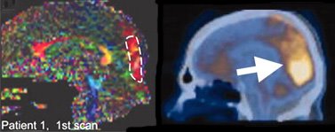
 The figure above illustrates Mr. Wallis' brain. On the left is a DTI scan to trace fiber tracts. On the right is a PET scan to measure glucose metabolism. The areas highlighted are on the posterior medial surface of the brain, the parietal-occipital region, which includes the cuneus and precuneus.
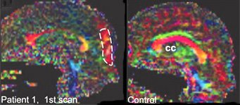
It's really interesting to compare the DTI scan of Mr. Wallis with that of a control subject, as illustrated below. The first thing to note is the degeneration in Patient 1's corpus callosum (the big red crescent in the middle of the control's brain, labeled cc). This structure is a huge white matter tract that connects the two cerebral hemispheres. But what's most novel is the unique white matter tract in Patient 1's parietal-occipital region. Could this presumed axonal growth be the basis of the remarkable improvement in Mr. Wallis' state of awareness?

 photo: Ron Phillips for The New York Times
- Finding Consciousness Within
It’s difficult to imagine anything worse than lying paralysed, being fully aware and yet unable to signal to your loved ones sitting around you that – yes, you can hear them, you are there. If only the doctors could scan your brain and see that you...
- Synesthetes Show Greater Connectivity In The Inferior Temporal Cortex
Ah ah Something ain't right I'm gonna get myself I'm gonna get myself I'm gonna get myself connected --Stereo MC's, Connected SYNESTHESIA can be defined as ...an unusual conscious experience, in which stimulation of one sensory modality...
- Daydreaming And Thought-sampling
OK, is there anything new in the daydreaming article in Science? Fig. 2. Graphs depict regions that exhibited a significant positive relation [with a propensity to daydream], r(14) > 0.50, P < .05 (A) Bilateral mPFC; (B) Bilateral precuneus and posterior...
- Are You Conscious Of Your Precuneus?
No, of course not. The question really is, does your precuneus make you conscious? In The Neurocritic's last entry on hypnosis and consciousness, Faymonville et al. (in press) raised the possibility that the precuneus "...is part of the critical neural...
- Deep Brain Stimulation
A curious case report discussed in today's New York Times: Man Regains Speech After Brain Stimulation By BENEDICT CAREY Published: August 1, 2007 A 38-year-old man who spent more than five years in a mute, barely conscious state as a result of a...
Neuroscience
The Precuneus and Recovery From a Minimally Conscious State
 You've probably heard about the case of Terry Wallis, a 42 year old man in Arkansas who spent 19 years in a "minimally conscious state" following severe traumatic brain injury. Then one day he saw his mother walk into the room and (after not speaking all those years) said "Mom."
The Aspen workgroup defined the minimally conscious state (MCS) as a condition of severely altered consciousness in which the person demonstrates minimal but definite behavioral evidence of self or environmental awareness (Giacino et al, 1997).A number of bloggers have commented on how his case is different from that of Terry Schiavo, who was in a persistent vegetative state (not a MCS).
What I'll focus on here is the work of researchers at Cornell, who followed Mr. Wallis using diffusion tensor imaging (DTI) and PET scanning over an 18 month period.
Using a novel technique, they saw evidence of new growth in the midline cerebellum, an area involved in motor control, as Mr. Wallis gained strength and range in his limbs. Another area of new growth, located along the back of the brain, is believed by some experts to be a central switching center for conscious awareness.The "central switching center for conscious awareness" is the precuneus, interestingly enough.
The daily exercises, the interactions with his parents, his regular dose of antidepressant medication: any or all of these might have spurred brain cells to grow more connections, the researchers said.
"The big missed opportunity is that we didn't know this guy would spontaneously emerge, and we didn't get to monitor him before then" to find out what preceded it, Dr. Schiff said.
Henning U. Voss, Aziz M. Uluç, Jonathan P. Dyke, Richard Watts, Erik J. Kobylarz, Bruce D. McCandliss, Linda A. Heier, Bradley J. Beattie, Klaus A. Hamacher, Shankar Vallabhajosula, Stanley J. Goldsmith, Douglas Ballon, Joseph T. Giacino and Nicholas D. Schiff. (2006). Possible axonal regrowth in late recovery from the minimally conscious state. J. Clin. Invest. 116: 2005-2011. OPEN ACCESS ARTICLE!In a commentary on the article, Laureys, Boly, and Maquet note the importance of the precuneus in conscious awareness:
We used diffusion tensor imaging (DTI) to study 2 patients with traumatic brain injury. The first patient recovered reliable expressive language after 19 years in a minimally conscious state (MCS); the second had remained in MCS for 6 years. Comparison of white matter integrity in the patients and 20 normal subjects using histograms of apparent diffusion constants and diffusion anisotropy identified widespread altered diffusivity and decreased anisotropy in the damaged white matter. These findings remained unchanged over an 18-month interval between 2 studies in the first patient. In addition, in this patient, we identified large, bilateral regions of posterior white matter with significantly increased anisotropy that reduced over 18 months. In contrast, notable increases in anisotropy within the midline cerebellar white matter in the second study correlated with marked clinical improvements in motor functions. This finding was further correlated with an increase in resting metabolism measured by PET in this subregion. Aberrant white matter structures were evident in the second patient’s DTI images but were not clinically correlated. We propose that axonal regrowth may underlie these findings and provide a biological mechanism for late recovery. Our results are discussed in the context of recent experimental studies that support this inference.
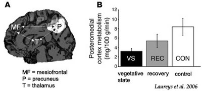
The most remarkable finding in the Voss et al. study (12) was the MRI assessment of transiently increased fractional anisotropy and directionality in the posterior midline cortices (encompassing the cuneus and precuneus), interpreted as increased myelinated fiber densities and novel corticocortical sprouting, paralleling the emergence of the patient from MCS. The same area of the patient’s brain also showed amplified metabolic activity, as measured by PET. This finding stresses the importance of the posterior medial structures in consciousness of self and interaction with the environment (14, 15). Activity in the medial parietal cortex (i.e., precuneus) seems to show it to be the brain region that best differentiates MCS from VS patients (16). Interestingly, this area is among the most active brain regions in conscious waking (15) and is among the least active in altered states of consciousness, such as pharmacological coma (17), sleep (18), dementia (19), Wernicke-Korsakoff syndrome, and postanoxic amnesia (20). It has been suggested that this richly connected multimodal posteromedial associative area is part of the neural network subserving human awareness (21).
 The figure above illustrates Mr. Wallis' brain. On the left is a DTI scan to trace fiber tracts. On the right is a PET scan to measure glucose metabolism. The areas highlighted are on the posterior medial surface of the brain, the parietal-occipital region, which includes the cuneus and precuneus.
It's really interesting to compare the DTI scan of Mr. Wallis with that of a control subject, as illustrated below. The first thing to note is the degeneration in Patient 1's corpus callosum (the big red crescent in the middle of the control's brain, labeled cc). This structure is a huge white matter tract that connects the two cerebral hemispheres. But what's most novel is the unique white matter tract in Patient 1's parietal-occipital region. Could this presumed axonal growth be the basis of the remarkable improvement in Mr. Wallis' state of awareness?

Steven Laureys, Mélanie Boly and Pierre Maquet (2006). Tracking the recovery of consciousness from coma. J. Clin. Invest. 116:1823-1825. OPEN ACCESS ARTICLE!
Predicting the chances of recovery of consciousness and communication in patients who survive their coma but transit in a vegetative state or minimally conscious state (MCS) remains a major challenge for their medical caregivers. Very few studies have examined the slow neuronal changes underlying functional recovery of consciousness from severe chronic brain damage. A case study in this issue of the JCI reports an extraordinary recovery of functional verbal communication and motor function in a patient who remained in MCS for 19 years (see the related article beginning on page 2005). Diffusion tensor MRI showed increased fractional anisotropy (assumed to reflect myelinated fiber density) in posteromedial cortices, encompassing cuneus and precuneus. These same areas showed increased glucose metabolism as studied by PET scanning, likely reflecting the neuronal regrowth paralleling the patient’s clinical recovery. This case shows that old dogmas need to be oppugned, as recovery with meaningful reduction in disability continued in this case for nearly 2 decades after extremely severe traumatic brain injury.
 photo: Ron Phillips for The New York Times
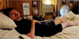
Terry Wallis at his home in Arkansas.
Mute 19 Years, He Helps Reveal Brain's Mysteries
. . .
He does not feel any physical pain, he told his parents, and he has no real sense of time. He also said recently that he was "proud" to be alive.
"It is good to know all that," said his father, sitting on the porch on Saturday evening.
"It's good to hear him say that, because if he didn't say so, you'd just have no way to know."
- Finding Consciousness Within
It’s difficult to imagine anything worse than lying paralysed, being fully aware and yet unable to signal to your loved ones sitting around you that – yes, you can hear them, you are there. If only the doctors could scan your brain and see that you...
- Synesthetes Show Greater Connectivity In The Inferior Temporal Cortex
Ah ah Something ain't right I'm gonna get myself I'm gonna get myself I'm gonna get myself connected --Stereo MC's, Connected SYNESTHESIA can be defined as ...an unusual conscious experience, in which stimulation of one sensory modality...
- Daydreaming And Thought-sampling
OK, is there anything new in the daydreaming article in Science? Fig. 2. Graphs depict regions that exhibited a significant positive relation [with a propensity to daydream], r(14) > 0.50, P < .05 (A) Bilateral mPFC; (B) Bilateral precuneus and posterior...
- Are You Conscious Of Your Precuneus?
No, of course not. The question really is, does your precuneus make you conscious? In The Neurocritic's last entry on hypnosis and consciousness, Faymonville et al. (in press) raised the possibility that the precuneus "...is part of the critical neural...
- Deep Brain Stimulation
A curious case report discussed in today's New York Times: Man Regains Speech After Brain Stimulation By BENEDICT CAREY Published: August 1, 2007 A 38-year-old man who spent more than five years in a mute, barely conscious state as a result of a...
