Neuroscience
No, of course not. The question really is, does your precuneus make you conscious?
In The Neurocritic's last entry on hypnosis and consciousness, Faymonville et al. (in press) raised the possibility that the precuneus "...is part of the critical neural network subserving conscious experience."
The second big boost for precuneus fans (and the one relevant for hypnosis) came from the aforementioned "default mode" studies of Raichle and colleagues (Gusnard & Raichle, 2001). The precuneus shows shows the highest resting metabolic rate of all the regions implicated in "the resting state" (often assessed with eyes closed, when the subjects presumably have a major alpha rhythm going in their EEGs, but also evaluated during "passive viewing" conditions when people are looking at a + sign or some other image). SO the hypothesis of these investigators (as quoted in Faymonville et al., in press) is that:
One of the most intriguing recent proposals was a speculation by Buckner and colleagues (2005), when pondering the meaning of an underactive "default mode" network in patients with Alzheimer's disease:
References
Buckner RL, Snyder AZ, Shannon BJ, LaRossa G, Sachs R, Fotenos AF, Sheline YI, Klunk WE, Mathis CA, Morris JC, Mintun MA.Molecular, structural, and functional characterization of Alzheimer's disease: evidence for a relationship between default activity, amyloid, and memory. J Neurosci. 2005; 25: 7709-17.
Fletcher PC, Frith CD, Baker SC, Shallice T, Frackowiak RS, Dolan RJ. The mind’s eye - precuneus activation in memory-related imagery. Neuroimage 1995; 2: 195-200.
Gusnard DA, Raichle ME. Searching for a baseline: functional imaging and the resting human brain. [Review]. Nat Rev Neurosci 2001; 2: 685-94.
Kennedy DP, Redcay E, Courchesne E. Failing to deactivate: resting functional abnormalities in autism. Proc Natl Acad Sci U S A. 2006; 103: 8275-80.
Krause BJ, Schmidt D, Mottaghy FM, Taylor J, Halsband U, Herzog H, et al. Episodic retrieval activates the precuneus irrespective of the imagery content of word pair associates: a PET study. Brain 1999; 122: 255-63.
NOTE: I know I promised you pain control last time, and that's still on The Neurocritic's busy agenda, which is filled with work-related obligations that interfere with blogging. Pah!
- Resisting A Resting State
The most forceful (and the only one published, it seems) objection to the concept of a "default mode" of processing in the human brain has been articulated by Alexa Morcom and Paul Fletcher at Cambridge. The brain's "default mode" or "resting state"...
- Daydreaming And Thought-sampling
OK, is there anything new in the daydreaming article in Science? Fig. 2. Graphs depict regions that exhibited a significant positive relation [with a propensity to daydream], r(14) > 0.50, P < .05 (A) Bilateral mPFC; (B) Bilateral precuneus and posterior...
- Hypnosis And Consciousness
Next in a continuing series on hypnosis: Functional neuroanatomy of the hypnotic state In Press, Corrected Proof, Journal of Physiology (Paris). Marie-Elisabeth Faymonville, Mélanie Boly and Steven Laureys What is hypnosis and how to induce it There...
- I Suggest... Neuroimaging Studies Of Hypnosis
Now back to our irregularly scheduled neuroscience programming! There's a series of in press articles about hypnosis in the Journal of Physiology (Paris). I'll take a look and report back. Functional neuroanatomy of the hypnotic state In...
- Neuropsychology Abstract Of The Day: Hippocampal Function And Alzheimer's Disease
Hippocampal hyperactivation associated with cortical thinning in Alzheimer's disease signature regions in non-demented elderly adults Journal of Neuroscience. 2011 Nov 30; 31(48): 17680-17688 Putcha D, Brickhouse M, O'Keefe K, Sullivan C, Rentz...
Neuroscience
Are You Conscious of Your Precuneus?
No, of course not. The question really is, does your precuneus make you conscious?
In The Neurocritic's last entry on hypnosis and consciousness, Faymonville et al. (in press) raised the possibility that the precuneus "...is part of the critical neural network subserving conscious experience."

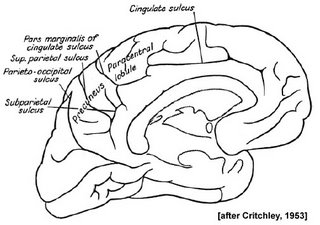
Andrea E. Cavanna and Michael R. Trimble (2006). The precuneus: a review of its functional anatomy and behavioural correlates. Brain 129: 564-583.The precuneus seemed poised to break out into the mainstream in the mid-90's, ever since it was found to be highly active when people were remembering words from a list they had studied earlier (compared to new words they hadn't studied). At first, this activity seemed related to the use of mental imagery during retrieval (Fletcher et al., 1995), because precuneus activity increased for highly concrete and imageable word pairs (e.g., "River-Stream") but not for abstract word pairs (e.g., "Justice-Law"). Subsequent experiments, however, did not replicate this result (Krause et al., 1999).
Functional neuroimaging studies have started unravelling unexpected functional attributes for the posteromedial portion of the parietal lobe, the precuneus. This cortical area has traditionally received little attention, mainly because of its hidden location and the virtual absence of focal lesion studies. However, recent functional imaging findings in healthy subjects suggest a central role for the precuneus in a wide spectrum of highly integrated tasks, including visuo-spatial imagery, episodic memory retrieval and self-processing operations, namely first-person perspective taking and an experience of agency. Furthermore, precuneus and surrounding posteromedial areas are amongst the brain structures displaying the highest resting metabolic rates (hot spots) and are characterized by transient decreases in the tonic activity during engagement in non-self-referential goal-directed actions (default mode of brain function). Therefore, it has recently been proposed that precuneus is involved in the interwoven network of the neural correlates of self-consciousness, engaged in self-related mental representations during rest. This hypothesis is consistent with the selective hypometabolism in the posteromedial cortex reported in a wide range of altered conscious states, such as sleep, drug-induced anaesthesia and vegetative states. ... [The authors describe] preliminary evidence for a functional subdivision within the precuneus into an anterior region, involved in self-centred mental imagery strategies, and a posterior region, subserving successful episodic memory retrieval.
The second big boost for precuneus fans (and the one relevant for hypnosis) came from the aforementioned "default mode" studies of Raichle and colleagues (Gusnard & Raichle, 2001). The precuneus shows shows the highest resting metabolic rate of all the regions implicated in "the resting state" (often assessed with eyes closed, when the subjects presumably have a major alpha rhythm going in their EEGs, but also evaluated during "passive viewing" conditions when people are looking at a + sign or some other image). SO the hypothesis of these investigators (as quoted in Faymonville et al., in press) is that:
the precuneus and interconnected posterior cingulate and medial prefrontal cortices are engaged in continuous information gathering and representation of the self and external world (Gusnard and Raichle, 2001).The "self-representation" part appears to be off in autistic individuals (Kennedy et al., 2006), because they fail to "engage" the default mode network during rest, relative to an active cognitive task condition. But in the same vein as the critique of the "Lose Yourself" study, a relatively quiet precuneus don't necessarily mean you're not conscious, it can just reflect a focus on things other than yourself and your surroundings. So perhaps the precuneus and its network of friends contribute to our "self-conscious" state.
One of the most intriguing recent proposals was a speculation by Buckner and colleagues (2005), when pondering the meaning of an underactive "default mode" network in patients with Alzheimer's disease:
Alzheimer's disease (AD) and antecedent factors associated with AD were explored using amyloid imaging and unbiased measures of longitudinal atrophy in combination with reanalysis of previous metabolic and functional studies. In total, data from 764 participants were compared across five in vivo imaging methods. Convergence of effects was seen in posterior cortical regions, including posterior cingulate, retrosplenial, and lateral parietal cortex. These regions were active in default states in young adults and also showed amyloid deposition in older adults with AD. At early stages of AD progression, prominent atrophy and metabolic abnormalities emerged in these posterior cortical regions; atrophy in medial temporal regions was also observed. Event-related functional magnetic resonance imaging studies further revealed that these cortical regions are active during successful memory retrieval in young adults. One possibility is that lifetime cerebral metabolism associated with regionally specific default activity predisposes cortical regions to AD-related changes, including amyloid deposition, metabolic disruption, and atrophy. These cortical regions may be part of a network with the medial temporal lobe whose disruption contributes to memory impairment.So basically, the idea is that an overactive self-referential default-mode network burns itself out, somehow leading to the pathological state associated with amyloid deposition, neuronal death, severe brain atrophy, and the ultimate loss of self which occurs in that dreadful disease named after Alois Alzheimer.
References
Buckner RL, Snyder AZ, Shannon BJ, LaRossa G, Sachs R, Fotenos AF, Sheline YI, Klunk WE, Mathis CA, Morris JC, Mintun MA.Molecular, structural, and functional characterization of Alzheimer's disease: evidence for a relationship between default activity, amyloid, and memory. J Neurosci. 2005; 25: 7709-17.
Fletcher PC, Frith CD, Baker SC, Shallice T, Frackowiak RS, Dolan RJ. The mind’s eye - precuneus activation in memory-related imagery. Neuroimage 1995; 2: 195-200.
Gusnard DA, Raichle ME. Searching for a baseline: functional imaging and the resting human brain. [Review]. Nat Rev Neurosci 2001; 2: 685-94.
Kennedy DP, Redcay E, Courchesne E. Failing to deactivate: resting functional abnormalities in autism. Proc Natl Acad Sci U S A. 2006; 103: 8275-80.
Krause BJ, Schmidt D, Mottaghy FM, Taylor J, Halsband U, Herzog H, et al. Episodic retrieval activates the precuneus irrespective of the imagery content of word pair associates: a PET study. Brain 1999; 122: 255-63.
NOTE: I know I promised you pain control last time, and that's still on The Neurocritic's busy agenda, which is filled with work-related obligations that interfere with blogging. Pah!
- Resisting A Resting State
The most forceful (and the only one published, it seems) objection to the concept of a "default mode" of processing in the human brain has been articulated by Alexa Morcom and Paul Fletcher at Cambridge. The brain's "default mode" or "resting state"...
- Daydreaming And Thought-sampling
OK, is there anything new in the daydreaming article in Science? Fig. 2. Graphs depict regions that exhibited a significant positive relation [with a propensity to daydream], r(14) > 0.50, P < .05 (A) Bilateral mPFC; (B) Bilateral precuneus and posterior...
- Hypnosis And Consciousness
Next in a continuing series on hypnosis: Functional neuroanatomy of the hypnotic state In Press, Corrected Proof, Journal of Physiology (Paris). Marie-Elisabeth Faymonville, Mélanie Boly and Steven Laureys What is hypnosis and how to induce it There...
- I Suggest... Neuroimaging Studies Of Hypnosis
Now back to our irregularly scheduled neuroscience programming! There's a series of in press articles about hypnosis in the Journal of Physiology (Paris). I'll take a look and report back. Functional neuroanatomy of the hypnotic state In...
- Neuropsychology Abstract Of The Day: Hippocampal Function And Alzheimer's Disease
Hippocampal hyperactivation associated with cortical thinning in Alzheimer's disease signature regions in non-demented elderly adults Journal of Neuroscience. 2011 Nov 30; 31(48): 17680-17688 Putcha D, Brickhouse M, O'Keefe K, Sullivan C, Rentz...
