Neuroscience
More on spindle neurons (or von Economo neurons), the next new thing:
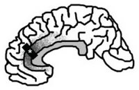
 The original paper, published in the Annals of Neurology (Seeley et al., 2006), used meticulous neuroanatomical quantification techniques to examine the neurons in layer V of the cerebral cortex from post-mortem brains with frontotemporal dementia, Alzheimer's disease, and neither disease (i.e., controls). The left pregenual anterior cingulate cortex (ACC) was the one region sampled (see figure). Due to tissue availability constraints, there were no samples from either right ACC or frontoinsular cortex from either hemisphere.
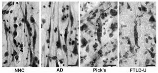
At any rate, the figure below illustrates the VENs in controls, Alzheimer's, Pick's disease, and frontotemporal dementia (aka frontotemporal lobar degeneration).

from Fig 3. Von Economo neuron (VEN) swelling and dysmorphism in frontotemporal dementia. VENs in nonneurological control subjects (NNC) and Alzheimer’s disease (AD) patients showed prominent clustering, smooth contours, and slender, tapering somata. In frontotemporal dementia, VENs were often solitary, swollen (especially in Pick’s disease), or showed twisting and kinking of proximal dendrites (both Pick’s and FTLD-U).
Unfortunately, media coverage of this finding has tended to overstate (or misstate) the significance and function of spindle neurons:
References
Nimchinsky EA, Vogt BA, Morrison JH, Hof PR. (1995). Spindle neurons of the human anterior cingulate cortex. J Comp Neurol 355: 27–37.
- Spindle Neurons In Elephants And Dolphins: Convergent Evolution In Large-brained Mammals?
Fig. 1 (Hakeem et al., 2009). Photomicrographs of VENs in the brain of the African elephant. A: VENs in frontoinsular cortex (area FI). Scale = 25 μm. Spindle neurons, or Von Economo neurons (VENs), are a unique type of large, bipolar neuron found in...
- Spindle Neurons And Science Writing
In the wake of a hyperbolic new article (Freedberg & Gallese, 2007) on trendoid mirror neurons, Neurobloggy Land has noted that spindle neurons (aka von Economo neurons) are competing for attention. In Neuroscience and Science Writing, Jonah at The...
- Spindle Neurons In Humpback Whales
from Hof & Van Der Gucht (2006) Spindle Neurons: The Next New Thing? In July, The Neurocritic wrote about a special class of cells found in the anterior cingulate cortex and frontoinsular cortex of humans and great apes. These spindle neurons are...
- Spindle Neurons: The Next New Thing?
In a neuroanatomical tour de force, Nimchinsky and colleagues (1999) obtained access to samples of the anterior cingulate cortex (and other cortical regions) from 28 different primate species, from prosimians to anthropoids to great apes to humans. They...
- Spindle Neurons
From the CBC: Humpback whales share brain cells with humans Last Updated: Monday, November 27, 2006 | 1:12 PM ET CBC News Humpback whales have joined an exclusive evolutionary club alongside humans, gorillas and dolphins, thanks to the discovery of a...
Neuroscience
Spindle Neurons and Frontotemporal Dementia
More on spindle neurons (or von Economo neurons), the next new thing:
Window into human behavior, brain disease
UCSF scientists have identified a cell population that is a primary target of the degenerative brain disease known as frontotemporal dementia, which is as common as Alzheimer's disease in patients who develop dementia before age 65.
Because the cells arose only recently in evolutionary history - in a common ancestor of great apes and humans - and are particularly abundant in humans, and the finding supports the concept that evolution has rendered the human brain vulnerable to disease, including frontotemporal dementia, and, possibly, disorders such as autism and schizophrenia, the researchers say.
In addition, because the disease erodes aspects of social behavior and emotions – self awareness, moral reasoning and empathy - that are highly developed in humans, the finding suggests that the cells may play a role in what makes humans "human," they say.
. . .
The cells received only limited attention in the ensuing years, but in the meantime scientists determined that the brain regions in which von Economo neurons arise - the anterior cingulate and frontoinsular cortex -- are key targets of frontotemporal dementia. And in 1999, a team of U.S. scientists made the surprising discovery that, among primates, von Economo neurons were seen only in great apes and humans.
 The original paper, published in the Annals of Neurology (Seeley et al., 2006), used meticulous neuroanatomical quantification techniques to examine the neurons in layer V of the cerebral cortex from post-mortem brains with frontotemporal dementia, Alzheimer's disease, and neither disease (i.e., controls). The left pregenual anterior cingulate cortex (ACC) was the one region sampled (see figure). Due to tissue availability constraints, there were no samples from either right ACC or frontoinsular cortex from either hemisphere.
Seeley WW, Carlin DA, Allman JM, Macedo MN, Bush C, Miller BL, Dearmond SJ. (2006). Early frontotemporal dementia targets neurons unique to apes and humans. Ann Neurol 60: 660-667.Oh, and here's a pretty important fact about von Economo neurons:
OBJECTIVE: Frontotemporal dementia (FTD) is a neurodegenerative disease that erodes uniquely human aspects of social behavior and emotion. The illness features a characteristic pattern of early injury to anterior cingulate and frontoinsular cortex. These regions, though often considered ancient in phylogeny, are the exclusive homes to the von Economo neuron (VEN), a large bipolar projection neuron found only in great apes and humans. Despite progress toward understanding the genetic and molecular bases of FTD, no class of selectively vulnerable neurons has been identified. METHODS: Using unbiased stereology, we quantified anterior cingulate VENs and neighboring Layer 5 neurons in FTD (n = 7), Alzheimer's disease (n = 5), and age-matched nonneurological control subjects (n = 7). Neuronal morphology and immunohistochemical staining patterns provided further information about VEN susceptibility. RESULTS: FTD was associated with early, severe, and selective VEN losses, including a 74% reduction in VENs per section compared with control subjects. VEN dropout was not attributable to general neuronal loss and was seen across FTD pathological subtypes. Surviving VENs were often dysmorphic, with pathological tau protein accumulation in Pick's disease. In contrast, patients with Alzheimer's disease showed normal VEN counts and morphology despite extensive local neurofibrillary pathology. INTERPRETATION: VEN loss links FTD to its signature regional pattern. The findings suggest a new framework for understanding how evolution may have rendered the human brain vulnerable to specific forms of degenerative illness.
VENs are thought to make up 1 to 2% of the Layer 5 neurons in ACC (Nimchinsky et al., 1995).So can 1-2% of cells in one layer (out of six total) of the cortex, with locations in only two circumscribed brain regions, mediate all of social behavior and emotions in humans (and great apes and humpback whales)? Sounds kind of preposterous when you put it that way.
At any rate, the figure below illustrates the VENs in controls, Alzheimer's, Pick's disease, and frontotemporal dementia (aka frontotemporal lobar degeneration).

from Fig 3. Von Economo neuron (VEN) swelling and dysmorphism in frontotemporal dementia. VENs in nonneurological control subjects (NNC) and Alzheimer’s disease (AD) patients showed prominent clustering, smooth contours, and slender, tapering somata. In frontotemporal dementia, VENs were often solitary, swollen (especially in Pick’s disease), or showed twisting and kinking of proximal dendrites (both Pick’s and FTLD-U).
Unfortunately, media coverage of this finding has tended to overstate (or misstate) the significance and function of spindle neurons:
'Social Dementia' Decimates Special NeuronsAbove link courtesy of Jake from Pure Pedantry. Check out his fine rant, There are no "social" neurons!
ScienceNOW Daily News
22 December 2006
. . .
Seeley and his colleagues conclude that VENs may play a key role in making humans the social creatures that we are, but that they also expose us to a higher risk of degenerative neural diseases. Lary Walker, a neuroscientist at Emory University in Atlanta, Georgia, says that the authors make a "reasonably compelling case that the VENs are selectively vulnerable in FTD". Nevertheless, Walker cautions against ascribing complex behaviors to the action of specific cells or regions in the brain.
References
Nimchinsky EA, Vogt BA, Morrison JH, Hof PR. (1995). Spindle neurons of the human anterior cingulate cortex. J Comp Neurol 355: 27–37.
- Spindle Neurons In Elephants And Dolphins: Convergent Evolution In Large-brained Mammals?
Fig. 1 (Hakeem et al., 2009). Photomicrographs of VENs in the brain of the African elephant. A: VENs in frontoinsular cortex (area FI). Scale = 25 μm. Spindle neurons, or Von Economo neurons (VENs), are a unique type of large, bipolar neuron found in...
- Spindle Neurons And Science Writing
In the wake of a hyperbolic new article (Freedberg & Gallese, 2007) on trendoid mirror neurons, Neurobloggy Land has noted that spindle neurons (aka von Economo neurons) are competing for attention. In Neuroscience and Science Writing, Jonah at The...
- Spindle Neurons In Humpback Whales
from Hof & Van Der Gucht (2006) Spindle Neurons: The Next New Thing? In July, The Neurocritic wrote about a special class of cells found in the anterior cingulate cortex and frontoinsular cortex of humans and great apes. These spindle neurons are...
- Spindle Neurons: The Next New Thing?
In a neuroanatomical tour de force, Nimchinsky and colleagues (1999) obtained access to samples of the anterior cingulate cortex (and other cortical regions) from 28 different primate species, from prosimians to anthropoids to great apes to humans. They...
- Spindle Neurons
From the CBC: Humpback whales share brain cells with humans Last Updated: Monday, November 27, 2006 | 1:12 PM ET CBC News Humpback whales have joined an exclusive evolutionary club alongside humans, gorillas and dolphins, thanks to the discovery of a...
