Neuroscience

The Agony of Genetically Disrupted Melanocortin Receptors (MC4R).
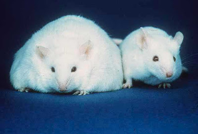
A new study suggests that blocking MC4R function in the central nervous system of rodents produces obesity by altering lipid metabolism and promoting fat deposition (Nogueiras et al., 2007). This effect was independent of food intake, suggesting a possible genetic propensity for obesity in humans (at least in a small percentage of the population). Wikipedia notes that

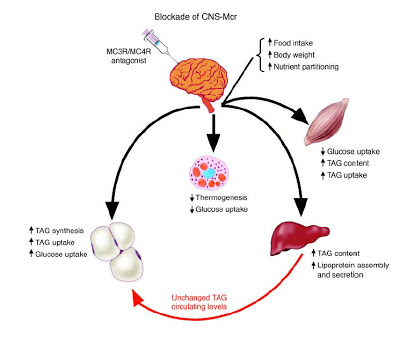
Figure 8 (Nogueiras et al., 2007). Schematic overview summarizing the physiological effects of CNS-Mcr blockade on peripheral tissues. Blockade of CNSMcr decreases thermogenesis and glucose utilization in BAT [brown adipose tissue]; decreases glucose utilization in muscle; increases TAG [triglyceride] content, lipoprotein assembly, and secretion in liver; and increases TAG synthesis and uptake as well as glucose uptake and insulin sensitivity in fat tissue. Combined, these parallel metabolic changes in multiple tissues represent a synergistic shift in substrate choice and nutrient partitioning, resulting in increased energy storage.
References
Adan RA, Tiesjema B, Hillebrand JJ, la Fleur SE, Kas MJ, de Krom M. (2006). The MC4 receptor and control of appetite. Br J Pharmacol. 149:815-27.
Farooqi IS, Keogh JM, Yeo GS, Lank EJ, Cheetham T, O'Rahilly S (2003). Clinical spectrum of obesity and mutations in the melanocortin 4 receptor gene. N. Engl. J. Med. 348:1085-95.
Nogueiras R, Wiedmer P, Perez-Tilve D, Veyrat-Durebex C, Keogh JM, Sutton GM, Pfluger PT, Castaneda TR, Neschen S, Hofmann SM, Howles PN, Morgan DA, Benoit SC, Szanto I, Schrott B, Schurmann A, Joost HG, Hammond C, Hui DY, Woods SC, Rahmouni K, Butler AA, Farooqi IS, O'rahilly S, Rohner-Jeanrenaud F, Tschop MH. (2007). The central melanocortin system directly controls peripheral lipid metabolism. J Clin Invest. 2007 Sep 20; [Epub ahead of print].
Disruptions of the melanocortin signaling system have been linked to obesity. We investigated a possible role of the central nervous melanocortin system (CNS-Mcr) in the control of adiposity through effects on nutrient partitioning and cellular lipid metabolism independent of nutrient intake. We report that pharmacological inhibition of melanocortin receptors (Mcr) in rats and genetic disruption of Mc4r in mice directly and potently promoted lipid uptake, triglyceride synthesis, and fat accumulation in white adipose tissue (WAT), while increased CNS-Mcr signaling triggered lipid mobilization. These effects were independent of food intake and preceded changes in adiposity. In addition, decreased CNS-Mcr signaling promoted increased insulin sensitivity and glucose uptake in WAT while decreasing glucose utilization in muscle and brown adipose tissue. Such CNS control of peripheral nutrient partitioning depended on sympathetic nervous system function and was enhanced by synergistic effects on liver triglyceride synthesis. Our findings offer an explanation for enhanced adiposity resulting from decreased melanocortin signaling, even in the absence of hyperphagia, and are consistent with feeding-independent changes in substrate utilization as reflected by respiratory quotient, which is increased with chronic Mcr blockade in rodents and in humans with loss-of-function mutations in MC4R. We also reveal molecular underpinnings for direct control of the CNS-Mcr over lipid metabolism. These results suggest ways to design more efficient pharmacological methods for controlling adiposity.
Obscurely Self-Referential
Obscure reference #1: I'm Not As Slim As That Girl
Obscure reference #2: The Joy Of Melanocortin Receptors
- Was I Wrong?
In honor of The Neurocritic's 10th anniversary, I'd like to announce a new occasional feature: Was I Wrong? In science, as in life, we learn from our mistakes. We can't move forward if we don't admit we were wrong and revise our entrenched...
- The Hunger:
The PYY and Dopamine Issue Brain 'hunger pathways' pinpointed 12:05 15 October 2007 Anna Gosline The brain circuitry that influences how much food a person will eat – whether they feel starving or full – has been revealed by a new imaging...
- The Joy Of 5-ht4 Receptors
Move over, melanocortin receptors, 5-HT4 receptors are ready to take your place. Agonists of the 5-HT4 receptor, a subtype of the serotonin receptor family, seem to do the following, according to recently published reports: (1) mediate the appetite-suppressing...
- Nip/tuck/npy Injections
"Tell me what you don't like about yourself." Scientists discover key to manipulating fat Washington, D.C. − In what they call a "stunning research advance," investigators at Georgetown University Medical Center have been able to use simple, non-toxic...
- Today's Disorder: Prader Willi Syndrome
According to the Prader-Willi Syndrome Association (UK): Prader-Willi Syndrome (PWS) was first described in 1956 by Swiss doctors, Prof. A Prader, Dr A Labhart and Dr H Willi, who recognised the condition as having unique and clearly definable features....
Neuroscience
I'm Not As Slim As That Mouse

The Agony of Genetically Disrupted Melanocortin Receptors (MC4R).
A new study suggests that blocking MC4R function in the central nervous system of rodents produces obesity by altering lipid metabolism and promoting fat deposition (Nogueiras et al., 2007). This effect was independent of food intake, suggesting a possible genetic propensity for obesity in humans (at least in a small percentage of the population). Wikipedia notes that
Defects in MC4R are a cause of autosomal dominant obesity, accounting for 6% of all cases of early-onset obesity (Farooqi et al., 2003).Conversely, stimulation of MC4R increases lipid mobilization (Nogueires et al., 2007) and reduces food intake (Adan et al., 2006). Thus,
Brain clue could provide anti-obesity drugsThe paper by Nogueiras et al. (PDF freely available) contains this wonderful diagram of how blocking melanocortin receptors affects fat storage and metabolism.
... Experiments on rats have shown that a part of the brain called the hypothalamus helps determine how much food energy is stored, raising the possibility of a new kind of anti-obesity drug.
Matthias Tschöp of the University of Cincinnati in Ohio used drugs that either stimulated or blocked receptors for the hormone melanocortin on hypothalamus cells in the brains of rats. Those given stimulatory drugs burned more of the carbohydrates in food, while those given inhibitory drugs converted them to fat and made extra fat in their liver (The Journal of Clinical Investigation).
Tschöp thinks the receptors communicate with fat and liver cells through the sympathetic nervous system, which also controls heartbeat and digestion. The same system may exist in humans, he believes, because people with faulty melanocortin receptors are often morbidly obese.

Figure 8 (Nogueiras et al., 2007). Schematic overview summarizing the physiological effects of CNS-Mcr blockade on peripheral tissues. Blockade of CNSMcr decreases thermogenesis and glucose utilization in BAT [brown adipose tissue]; decreases glucose utilization in muscle; increases TAG [triglyceride] content, lipoprotein assembly, and secretion in liver; and increases TAG synthesis and uptake as well as glucose uptake and insulin sensitivity in fat tissue. Combined, these parallel metabolic changes in multiple tissues represent a synergistic shift in substrate choice and nutrient partitioning, resulting in increased energy storage.
References
Adan RA, Tiesjema B, Hillebrand JJ, la Fleur SE, Kas MJ, de Krom M. (2006). The MC4 receptor and control of appetite. Br J Pharmacol. 149:815-27.
Farooqi IS, Keogh JM, Yeo GS, Lank EJ, Cheetham T, O'Rahilly S (2003). Clinical spectrum of obesity and mutations in the melanocortin 4 receptor gene. N. Engl. J. Med. 348:1085-95.
Nogueiras R, Wiedmer P, Perez-Tilve D, Veyrat-Durebex C, Keogh JM, Sutton GM, Pfluger PT, Castaneda TR, Neschen S, Hofmann SM, Howles PN, Morgan DA, Benoit SC, Szanto I, Schrott B, Schurmann A, Joost HG, Hammond C, Hui DY, Woods SC, Rahmouni K, Butler AA, Farooqi IS, O'rahilly S, Rohner-Jeanrenaud F, Tschop MH. (2007). The central melanocortin system directly controls peripheral lipid metabolism. J Clin Invest. 2007 Sep 20; [Epub ahead of print].
Disruptions of the melanocortin signaling system have been linked to obesity. We investigated a possible role of the central nervous melanocortin system (CNS-Mcr) in the control of adiposity through effects on nutrient partitioning and cellular lipid metabolism independent of nutrient intake. We report that pharmacological inhibition of melanocortin receptors (Mcr) in rats and genetic disruption of Mc4r in mice directly and potently promoted lipid uptake, triglyceride synthesis, and fat accumulation in white adipose tissue (WAT), while increased CNS-Mcr signaling triggered lipid mobilization. These effects were independent of food intake and preceded changes in adiposity. In addition, decreased CNS-Mcr signaling promoted increased insulin sensitivity and glucose uptake in WAT while decreasing glucose utilization in muscle and brown adipose tissue. Such CNS control of peripheral nutrient partitioning depended on sympathetic nervous system function and was enhanced by synergistic effects on liver triglyceride synthesis. Our findings offer an explanation for enhanced adiposity resulting from decreased melanocortin signaling, even in the absence of hyperphagia, and are consistent with feeding-independent changes in substrate utilization as reflected by respiratory quotient, which is increased with chronic Mcr blockade in rodents and in humans with loss-of-function mutations in MC4R. We also reveal molecular underpinnings for direct control of the CNS-Mcr over lipid metabolism. These results suggest ways to design more efficient pharmacological methods for controlling adiposity.
Obscurely Self-Referential
Obscure reference #1: I'm Not As Slim As That Girl
Obscure reference #2: The Joy Of Melanocortin Receptors
- Was I Wrong?
In honor of The Neurocritic's 10th anniversary, I'd like to announce a new occasional feature: Was I Wrong? In science, as in life, we learn from our mistakes. We can't move forward if we don't admit we were wrong and revise our entrenched...
- The Hunger:
The PYY and Dopamine Issue Brain 'hunger pathways' pinpointed 12:05 15 October 2007 Anna Gosline The brain circuitry that influences how much food a person will eat – whether they feel starving or full – has been revealed by a new imaging...
- The Joy Of 5-ht4 Receptors
Move over, melanocortin receptors, 5-HT4 receptors are ready to take your place. Agonists of the 5-HT4 receptor, a subtype of the serotonin receptor family, seem to do the following, according to recently published reports: (1) mediate the appetite-suppressing...
- Nip/tuck/npy Injections
"Tell me what you don't like about yourself." Scientists discover key to manipulating fat Washington, D.C. − In what they call a "stunning research advance," investigators at Georgetown University Medical Center have been able to use simple, non-toxic...
- Today's Disorder: Prader Willi Syndrome
According to the Prader-Willi Syndrome Association (UK): Prader-Willi Syndrome (PWS) was first described in 1956 by Swiss doctors, Prof. A Prader, Dr A Labhart and Dr H Willi, who recognised the condition as having unique and clearly definable features....
