Neuroscience
 In an interview with ReachMD, John Y.K. Lee, MD, medical director of the Penn Gamma Knife Center and assistant professor of neurosurgery, discusses 3-D endoscopy and its advantages over 2-D technology in the removal of brain tumors.
When surgeons use their own eyes to perform surgery, they are operating in “3-D” because the naked eye produces and delivers a three-dimensional image to the brain. As minimally invasive surgical techniques have replaced open surgery as the preferred treatment for more and more conditions, surgeons, in turn, increasingly rely on tiny “endoscopes” to treat many hard-to-reach places in the body.
The endoscope captures and transmits images to the physician from inside the body. In many cases, a minimally invasive endoscopy uses a natural orifice. Neurosurgeons perform endoscopic brain surgery using the nose as an access point to the brain.
While 3-D endoscopy technology for other areas of the body has existed for some time, neurosurgeons have only recently seen the development of three-dimensional endoscopes. “3-D technology has been in use for robotic surgery in other areas of the body for years—the three-dimensional view is achieved by putting two endoscopes side-by-side. The problem for neurosurgery was that these two endoscopes together are too big to fit through the nose—it didn’t work for brain surgery,” says Dr. Lee.
New neurosurgical endoscopes provide a 3-D image from a single endoscope and are used to remove certain types of brain tumors. Where a 2-D endoscope uses fiber optics to deliver images outside the body to a camera, this new generation of 3-D endoscopes is equipped with a microprocessor computer chip at the end of the scope. This “chip tip” technology records imaging on-site in the body and transmits a crisp 3-D image to the physician on a 3-D television screen.
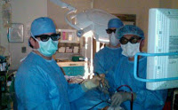
“We wear the [3-D] glasses just like you do when you watch a 3-D movie, but we use ‘polarized’ glasses which are lighter. This is important—especially for surgical cases that can last up to 10 hours,” notes Dr. Lee.
Endoscopic neurosurgery is ideal for ventral skull base tumors including:
• Pituitary tumor
• Meningioma
• Craniopharyngioma
• Clival chordoma
Neurosurgeons using a 2-D endoscope carefully navigate their way to the brain through the nose and remove the tumor using a number of techniques, including palpation (feeling the way through the nose as one gets closer and closer to the tumor) and motion parallax (moving the endoscope in and out, and comparing the different views for depth perception).
Dr. Lee describes the critical nature of endoscopic neurosurgery and the advantage of 3-D imaging, “The addition of the third dimension gives me real clinical and surgical benefit. When I’m operating near critical structures in the brain, if I move incorrectly, the patient could be blinded or have a stroke. With the 3-D technology, I can see the tumor and surrounding structures like the arteries, nerves and pituitary gland in crisp detail. And because this technology is based on microprocessor chips, the potential for advancement is exponential.”
Listen Now: ReachMD Interview with John Y.K. Lee, MD
- Five Surgeons From Penn Neurosurgery Earn 'top Doc' Honors
Each year, Philadelphia magazine recognizes the area’s outstanding doctors in their Top Doctors issue. The list is viewed as a “gold standard for those seeking the finest medical care in the Philadelphia area.” We are pleased to announce that five...
- Darren Daulton Diagnosed With 2 Brain Tumors
NBC10 reports that Darren Daulton, a beloved member of the Philadelphia Phillies for 14 seasons, has been diagnosed with two brain tumors. Penn neurosurgeon Donald O'Rourke, MD, says it depends on the location of the tumor, and not the size, for it...
- Five Surgeons From Penn Neurosurgery Earn 'top Doc' Honors
Each year, Philadelphia magazine recognizes the area’s outstanding doctors in their Top Doctors issue. The list is viewed as a “gold standard for those seeking the finest medical care in the Philadelphia area.” We are pleased to announce that five...
- Gamma Knife® Perfexion
Image courtesy of Elekta Gamma Knife® PerfexionTM is the latest form of radiation therapy that damages cancer cells and tumors, and prevents them from multiplying. It does not involve surgery, but rather precise beams of radiation delivered...
- John Y.k. Lee, Md, Receives Meningioma Mommas Grant Award
Meningiomas are the most common brain tumor in adults. However, they are often overlooked because they are considered a benign tumor. In many cases, meningiomas appear to be benign but are actually “atypical” and even malignant. These aggressive...
Neuroscience
3-D Endoscopic Brain Surgery

When surgeons use their own eyes to perform surgery, they are operating in “3-D” because the naked eye produces and delivers a three-dimensional image to the brain. As minimally invasive surgical techniques have replaced open surgery as the preferred treatment for more and more conditions, surgeons, in turn, increasingly rely on tiny “endoscopes” to treat many hard-to-reach places in the body.
The endoscope captures and transmits images to the physician from inside the body. In many cases, a minimally invasive endoscopy uses a natural orifice. Neurosurgeons perform endoscopic brain surgery using the nose as an access point to the brain.
While 3-D endoscopy technology for other areas of the body has existed for some time, neurosurgeons have only recently seen the development of three-dimensional endoscopes. “3-D technology has been in use for robotic surgery in other areas of the body for years—the three-dimensional view is achieved by putting two endoscopes side-by-side. The problem for neurosurgery was that these two endoscopes together are too big to fit through the nose—it didn’t work for brain surgery,” says Dr. Lee.
New neurosurgical endoscopes provide a 3-D image from a single endoscope and are used to remove certain types of brain tumors. Where a 2-D endoscope uses fiber optics to deliver images outside the body to a camera, this new generation of 3-D endoscopes is equipped with a microprocessor computer chip at the end of the scope. This “chip tip” technology records imaging on-site in the body and transmits a crisp 3-D image to the physician on a 3-D television screen.
“We wear the [3-D] glasses just like you do when you watch a 3-D movie, but we use ‘polarized’ glasses which are lighter. This is important—especially for surgical cases that can last up to 10 hours,” notes Dr. Lee.
Endoscopic neurosurgery is ideal for ventral skull base tumors including:
• Pituitary tumor
• Meningioma
• Craniopharyngioma
• Clival chordoma
Neurosurgeons using a 2-D endoscope carefully navigate their way to the brain through the nose and remove the tumor using a number of techniques, including palpation (feeling the way through the nose as one gets closer and closer to the tumor) and motion parallax (moving the endoscope in and out, and comparing the different views for depth perception).
Dr. Lee describes the critical nature of endoscopic neurosurgery and the advantage of 3-D imaging, “The addition of the third dimension gives me real clinical and surgical benefit. When I’m operating near critical structures in the brain, if I move incorrectly, the patient could be blinded or have a stroke. With the 3-D technology, I can see the tumor and surrounding structures like the arteries, nerves and pituitary gland in crisp detail. And because this technology is based on microprocessor chips, the potential for advancement is exponential.”
Listen Now: ReachMD Interview with John Y.K. Lee, MD
- Five Surgeons From Penn Neurosurgery Earn 'top Doc' Honors
Each year, Philadelphia magazine recognizes the area’s outstanding doctors in their Top Doctors issue. The list is viewed as a “gold standard for those seeking the finest medical care in the Philadelphia area.” We are pleased to announce that five...
- Darren Daulton Diagnosed With 2 Brain Tumors
NBC10 reports that Darren Daulton, a beloved member of the Philadelphia Phillies for 14 seasons, has been diagnosed with two brain tumors. Penn neurosurgeon Donald O'Rourke, MD, says it depends on the location of the tumor, and not the size, for it...
- Five Surgeons From Penn Neurosurgery Earn 'top Doc' Honors
Each year, Philadelphia magazine recognizes the area’s outstanding doctors in their Top Doctors issue. The list is viewed as a “gold standard for those seeking the finest medical care in the Philadelphia area.” We are pleased to announce that five...
- Gamma Knife® Perfexion
Image courtesy of Elekta Gamma Knife® PerfexionTM is the latest form of radiation therapy that damages cancer cells and tumors, and prevents them from multiplying. It does not involve surgery, but rather precise beams of radiation delivered...
- John Y.k. Lee, Md, Receives Meningioma Mommas Grant Award
Meningiomas are the most common brain tumor in adults. However, they are often overlooked because they are considered a benign tumor. In many cases, meningiomas appear to be benign but are actually “atypical” and even malignant. These aggressive...
