Self-Embedding Disorder and Removal of Soft Tissue Foreign Bodies

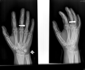
Figure 1 (Young et al., 2008). This x-ray image illustrates 3 metal staples embedded in the hand of a teenage girl.
Self-Embedding Disorder appears to be a newly-coined term1 described in a press release issued by the Radiological Society of North America (RSNA):
Radiologists Diagnose and Treat Self-Embedding Disorder in TeensIt's a form of self-injury, which has been widely reported in the literature (more broadly), and is well-known to clinicians.CHICAGO — Minimally invasive, image-guided treatment is a safe and precise method for removal of self-inflicted foreign objects from the body, according to the first report on "self-embedding disorder," or self-injury and self-inflicted foreign body insertion in adolescents. The findings will be presented today at the annual meeting of the Radiological Society of North America (RSNA).
"Radiologists are in a unique position to be the first to detect self-embedding disorder, make the appropriate diagnosis and mobilize the healthcare system for early and effective intervention and treatment," said the study's principal investigator, William E. Shiels II, D.O., chief of the Department of Radiology at Nationwide Children’s Hospital in Columbus, Ohio.
As the press release explains:
Self-injury, or self-harm, refers to a variety of behaviors in which a person intentionally inflicts harm to his or her body without suicidal intent. It is a disturbing trend among U.S. adolescents, particularly girls. Prevalence is unknown because many cases go unreported, but recent studies have reported that 13 to 24 percent of high school students in the U.S. and Canada have practiced deliberate self-injury at least once. More common forms of self-injury include cutting of the skin, burning, bruising, hair pulling, breaking bones or swallowing toxic substances. In cases of self-embedding disorder, objects are used to puncture the skin or are embedded into the wound after cutting.The interventional radiologists enter the scene (and intervene) when they use imaging to assist in the removal of self-inflicted soft tissue foreign bodies (STFBs). The abstract below (from an RNSA presentation on December 4, 2008) says it all...
Self-Mutilation in Adolescents: Radiological Management of Self-inflicted Soft Tissue Foreign Bodies
Adam Young, William Shiels, James Murakami, Brian Coley and Mark Hogan
PURPOSETo evaluate the efficacy and clinical impact of image-guided foreign body removal (IGFBR) for treatment of self-inflicted soft tissue foreign bodies (STFBs).
METHOD AND MATERIALSFour hundred patients underwent IGFBR with sonographic and/or fluoroscopic guidance. Self-mutilation was seen in 5 adolescent female patients (1.2%), representing 7 patient care encounters; 2 patients presented with self-inflicted STFBs on 2 separate occasions. Mean age 16.8 yr; (range 15-17 yr). Foreign body number, location, type and size as well as incision size, intraoperative imaging modality, type of surrounding reaction, and success or failure of removal were documented prospectively.
RESULTSTwenty-five foreign bodies were inserted into the forearm or upper arm of the five patients. Referring services included Pediatric Surgery, Emergency Department, and Psychiatry. Number of STFBs per case ranged from 1-9; median=2. Foreign body types included metal (13), wood (5), graphite (3), plastic (2), crayon (1), and stone (1). STFB measurement (greatest dimension) ranged from 4.5-160 mm; mean=22.06 mm. During sonographic removal, hypoechoic halos representing purulent material surrounding the STFBs were defined in 2 cases. Mean incision = 4.67 mm; STFBs were removed with sonographic guidance in 3 cases, fluoroscopic guidance in 3 cases, and a combination of the two modalities in 1 case. IGFBR was successful in all 7 cases without fragmentation or complications.
CONCLUSIONPercutaneous radiological treatment of self-inflicted STFBs is safe, precise, and effective for radiopaque and non-radiopaque foreign bodies, including foreign bodies at risk for fragmentation during traditional operative removal techniques.
CLINICAL RELEVANCE/APPLICATIONPercutaneous IGFBR with sonography and/or fluoroscopy offers surgeons and emergency physicians a safe and effective alternative to operative foreign body removal in this unique high-risk population.

Footnote
1 The term was not found in PubMed.
ADDENDUM: Psychologist Dr Lisa Boesky wants parents to understand that this is a very extreme disorder. "This is not new," she says. "I have been dealing with people who do this since 1995 in juvenile jails and prison. It's very rare in the public arena. Teens who embed typically have major mental health disorders and frequently have histories of severe sexual abuse or trauma."-via Momlogic.
ADDENDUM #2: The spelling error in the title [formerly ...Soft Tissue Foreign Bodes] has been corrected, thanks to psychiatrist Dr. Eliot Gelwan, who also notes, "...there is no need for a new diagnosis. Indeed, self-injuriousness in general is not an illness, or a diagnosis, unto itself, but rather a symptom of a variety of diagnoses. A fortiori for a particular kind of self-injuriousness. This illustrates one of the epistemological confusions plaguing the system for diagnosing behavioral problems, and is a perfect example of the needless proliferation of diagnostic categories."
- Developmental "foreign Accent Syndrome" - Cases Documented For The First Time
You may have seen cases of foreign accent syndrome (FAS) covered in the news. In 2007, for example, a ten-year-old boy acquired a new accent after undergoing brain surgery. "He went in with a York accent and came out all posh" his mother told the...
- Body Image - It's 'healthy' People Who Are Deluded
We’re all going to die and there’s nothing we can do about it. Depressing? Well, it’s been argued that depressed people are the sane ones because they see the world for how it really is. Now consider this – a study has found people with eating...
- Christmas Cheer From Bmj
Fig 1 (Firth et al., 2009). X ray pictures can easily detect an ingested coin. Position of coin on lateral view (left), relative to anterior (right) or posterior picture affects size of image on film. Every year, BMJ has a special Christmas issue with...
- Risperdal (risperidone) And Pediatric Schizophrenia And Bipolar Disorder
An FDA press release from yesterday: FDA Approves Risperdal for Two Psychiatric Conditions in Children and Adolescents The U.S. Food and Drug Administration today approved Risperdal (risperidone) for the treatment of schizophrenia in adolescents, ages...
- Mri During Brain Surgery
A press release from the Radiological Society of North America. Here is the abstract of the specific study mentioned in the release: [abstract]. MR imaging during brain surgery improves tumor removal OAK BROOK, Ill. - A specially adapted magnetic...
