Neuroscience

-Marcel Duchamp, Nude Descending a Staircase, No. 2
Neuroesthetics, a term coined by Semir Zeki, is "the attempt to use neuroscience to understand art and aesthetic behaviour" (as defined in an excellent overview by BRAINETHICS). Some would say the venture is anathema, that reductionistic explanations of the sublime are misguided at best and destructive at worst. Others hold that since all qualia emanate from the brain, a neuroscientific approach is the only true way to understand aesthetic experience.
Here's an early manifesto on neuroesthetics and visual art:

An earlier paper by Kawabata and Zeki (2004) studied the neural correlates of beauty. Prior to getting scanned,

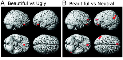
Adapted from Fig. 3 (Kawabata & Zeki, 2004). Statistical parametric maps rendered onto a standard brain showing judgment-specific activity in comparisons of (A) beautiful vs. ugly, and (B) beautiful vs. neutral. (A) shows medial orbito-frontal cortex only (Talairach coordinates –2, 36, –22). (B) medial orbito-frontal cortex (–2, 50, –20), anterior cingulate gyrus (–4, 48, 14) and left parietal cortex (–54, –68, 26).
In their study on beauty and pain, Tammaso and colleagues used the aesthetic rating methods of Kawabata and Zeki (2004) to classify the set of paintings for each participant. Several days after the rating task, the 12 subjects participated in an EEG experiment. Brain waves were recorded while they not only viewed the paintings, but also while a laser delivered unpleasantly hot stimuli to their hands.

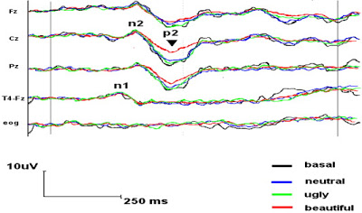
Fig. 3 (Tommaso et al., 2008). Grand average of LEPs obtained by stimulation of the left hand in the different experimental phases. The Fz, Cz, Pz and contralateral temporal (T4) recordings are shown. Compared with the baseline condition, the amplitude of the vertex N2–P2 component is smaller while viewing beautiful pictures. No changes were observed in the middle-latency N1 amplitude throughout the experiment.
The authors concluded that
Footnote
1 Although the activity of the ACC was increased in the beautiful vs. neutral contrast of Kawabata and Zeki but reduced by beauty during painful laser stimulation, the precise regions might be different in the two studies.
References
Kawabata H, Zeki S. (2004 ). Neural correlates of beauty. J Neurophysiol. 91:1699-705.
M TOMMASO, M SARDARO, P LIVREA (2008). Aesthetic value of paintings affects pain thresholds. Consciousness and Cognition DOI: 10.1016/j.concog.2008.07.002
Zeki S, Lamb M. (1994). The neurology of kinetic art. Brain 117:607-36.
- Chris Mcmanus: Beauty
What is this thing I call beauty? Not "art" as a social phenomenon based on status or display, or beautiful faces seen merely as biological fitness markers. Rather, the sheer, drawing-in-of-breath beauty of a Handel aria, a Rothko painting, TS Eliot’s...
- Imaging The Brain To Control The Mind
For the first time anywhere in the world, psychologists at California-based company Omneuron and Stanford University have demonstrated that people can be taught how to reduce their experience of pain with the aid of real-time images of their brain activity....
- The Neurophysiology Of Pain During Rem Sleep
In the last post, we learned about The Phenomenology of Pain During REM Sleep. Real life pain can intrude into dreams, as was shown for experimentally induced pain (Nielsen et al., 1993) and in hospitalized burn patients (Raymond et al., 2002). In this...
- Does It Look Painful Or Disgusting? Ask Your Parietal And Cingulate Cortex
...............Ouch!............Yuck! Figure 1 (Benuzzi et al., 2008). Sample frames extracted from some video clips representing painful (left), disgusting (middle), and neutral (right) stimuli. All video clips began with 200–400 ms of a static hand...
- Humor, Hot Flashes, And Empathy For Pain
These three phenomena activate the same brain areas (anterior cingulate cortex and frontoinsular cortex), according to recent findings. Any theory about the neural correlates of empathy must take into account the fact that the same brain regions are activated...
Neuroscience
Pain & Paintings: Beholding Beauty Reduces Pain Perception and Laser Evoked Potentials

-Marcel Duchamp, Nude Descending a Staircase, No. 2
Neuroesthetics, a term coined by Semir Zeki, is "the attempt to use neuroscience to understand art and aesthetic behaviour" (as defined in an excellent overview by BRAINETHICS). Some would say the venture is anathema, that reductionistic explanations of the sublime are misguided at best and destructive at worst. Others hold that since all qualia emanate from the brain, a neuroscientific approach is the only true way to understand aesthetic experience.
Here's an early manifesto on neuroesthetics and visual art:
Credo (manifesto of physiological facts)The above passage serves as background to an exploration of how visual art can literally affect our bodies. A new paper by Tommaso et al. (2008) used the methods of neuroesthetics to examine whether the act of viewing beautiful art (as individually defined by each participant) would reduce the perception of pain as well as its physiological manifestations.
All visual art must obey the laws of the visual system.The first law is that an image of the visual world is not impressed upon the retina, but assembled together in the visual cortex. Consequently, many of the visual phenomena traditionally attributed to the eye actually occur in the cortex. Among these is visual motion.The second law is that of the functional specialization of the visual cortex, by which we mean that separate attributes of the visual scene are processed in geographically separate parts of the visual cortex, before being combined to give a unified and coherent picture of the visual world.The third law is that the attributes that are separated, and separately processed, in the cerebral cortex are those which have primacy in vision. These are colour, form, motion and, possibly, depth. It follows that motion is an autonomous visual attribute, separately processed and therefore capable of being separately compromised after brain lesions. It is also one of the visual attributes that have primacy, just like form or colour or depth.We conclude that it is this separate visual attribute which those involved in kinetic art have tried to exploit, instinctively and physiologically, from which it follows that in their explorations artists are unknowingly exploring the organization of the visual brain though with techniques unique to them.
-S. Zeki and M. Lamb (1994), The Neurology of Kinetic Art (PDF).

An earlier paper by Kawabata and Zeki (2004) studied the neural correlates of beauty. Prior to getting scanned,
each subject viewed 300 paintings for each painting category that were reproductions viewed on a computer monitor. Each painting was given a score, on a scale from 1 to 10. Scores of 1–4 were classified as "ugly," 5–6 "neutral," and 7–10 "beautiful." Each subject thus arrived at an independent assessment of beautiful, ugly, and neutral paintings. Paintings classified as beautiful by some were classified as ugly by others and vice versa with the consequence that any individual painting did not necessarily belong in the same category for different subjects. Based on these psychophysical tests, a total of 16 stimuli in each category (abstract, still life, landscape, or portrait) were viewed by subjects in the scanner... However, in the ugly and beautiful categories, only paintings classified as 1–2 and 9–10, respectively, were viewed in the scanner, whereas for paintings classified as neutral, paintings belonging to both categories 5 and 6 were viewed.In the scanner, the participants performed the same aesthetic rating task. Paintings rated as beautiful were associated with greater activity in the medial orbitofrontal cortex (when compared to ugly or neutral paintings). Additional activations were observed in anterior cingulate cortex and left parietal cortex for the beautiful vs. neutral contrast.

Adapted from Fig. 3 (Kawabata & Zeki, 2004). Statistical parametric maps rendered onto a standard brain showing judgment-specific activity in comparisons of (A) beautiful vs. ugly, and (B) beautiful vs. neutral. (A) shows medial orbito-frontal cortex only (Talairach coordinates –2, 36, –22). (B) medial orbito-frontal cortex (–2, 50, –20), anterior cingulate gyrus (–4, 48, 14) and left parietal cortex (–54, –68, 26).
In their study on beauty and pain, Tammaso and colleagues used the aesthetic rating methods of Kawabata and Zeki (2004) to classify the set of paintings for each participant. Several days after the rating task, the 12 subjects participated in an EEG experiment. Brain waves were recorded while they not only viewed the paintings, but also while a laser delivered unpleasantly hot stimuli to their hands.
Cutaneous heat stimuli were delivered on the dorsum of the left hand. ... The location of impact on the skin was adjusted slightly between two successive stimuli to avoid nociceptor sensitization and fatigue. A laser (7.5 W) was used in each case, with a duration of 25 ms. The subjects were requested to rate the quality of sensation (pain rating: PR) after each series of stimuli on a visual analogue scale (VAS) where 0 indicated no pain in white, increasing in a gradual scale of reds to 100, which indicated the worst possible pain.So the subjects rated their level of pain on a visual analogue scale while looking at the various paintings and getting fried with a laser. Presentation of the painful stimuli evoked a specific type of EEG response, called laser evoked potentials (LEPs). A sequence of three LEPs were recorded in rapid succession, within the first 400 milliseconds after laser stimulation. These responses are called the N1, N2 and P2 potentials.
The first LEP component commonly identified is the N1, a negativity that is seen in temporal EEG-leads at about 150 ms following the laser stimulus (or later, depending on the type of laser). Its generator has been localized in the operculo–insular cortex, and this component has been demonstrated to be involved in focussed attention or spatial discrimination. The second is a large positive-negative complex named N2–P2 with its maximum amplitude at the vertex, generating from the posterior zone of the anterior cingulate cortex and expressing the variations in arousal and emotive reaction against pain.Remarkably, the P2 potential was smaller when participants were viewing beautiful paintings, and their subjective pain ratings were lower than in the other conditions (baseline, neutral, ugly). The figure below shows LEPs from electrodes placed on the scalp over frontal, central, and parietal midline regions, and over the right temporal lobe.

Fig. 3 (Tommaso et al., 2008). Grand average of LEPs obtained by stimulation of the left hand in the different experimental phases. The Fz, Cz, Pz and contralateral temporal (T4) recordings are shown. Compared with the baseline condition, the amplitude of the vertex N2–P2 component is smaller while viewing beautiful pictures. No changes were observed in the middle-latency N1 amplitude throughout the experiment.
The authors concluded that
aesthetic perception can affect subjective sensation and pain-related cortical responses. It is generally accepted that distraction produces analgesia (Johnson, 2005). The present study revealed that there is a different effect of distraction on pain depending on the type of distracting stimulus used. The vision of beautiful images seemed to have the maximum distractive effect from pain, as is shown by the lower subjective pain rating and the inhibition of pain-related evoked responses.Beauty is not only in the eye of the beholder, it modulates pain-related activity in the anterior cingulate cortex.1
Footnote
1 Although the activity of the ACC was increased in the beautiful vs. neutral contrast of Kawabata and Zeki but reduced by beauty during painful laser stimulation, the precise regions might be different in the two studies.
References
Kawabata H, Zeki S. (2004 ). Neural correlates of beauty. J Neurophysiol. 91:1699-705.
M TOMMASO, M SARDARO, P LIVREA (2008). Aesthetic value of paintings affects pain thresholds. Consciousness and Cognition DOI: 10.1016/j.concog.2008.07.002
Zeki S, Lamb M. (1994). The neurology of kinetic art. Brain 117:607-36.
- Chris Mcmanus: Beauty
What is this thing I call beauty? Not "art" as a social phenomenon based on status or display, or beautiful faces seen merely as biological fitness markers. Rather, the sheer, drawing-in-of-breath beauty of a Handel aria, a Rothko painting, TS Eliot’s...
- Imaging The Brain To Control The Mind
For the first time anywhere in the world, psychologists at California-based company Omneuron and Stanford University have demonstrated that people can be taught how to reduce their experience of pain with the aid of real-time images of their brain activity....
- The Neurophysiology Of Pain During Rem Sleep
In the last post, we learned about The Phenomenology of Pain During REM Sleep. Real life pain can intrude into dreams, as was shown for experimentally induced pain (Nielsen et al., 1993) and in hospitalized burn patients (Raymond et al., 2002). In this...
- Does It Look Painful Or Disgusting? Ask Your Parietal And Cingulate Cortex
...............Ouch!............Yuck! Figure 1 (Benuzzi et al., 2008). Sample frames extracted from some video clips representing painful (left), disgusting (middle), and neutral (right) stimuli. All video clips began with 200–400 ms of a static hand...
- Humor, Hot Flashes, And Empathy For Pain
These three phenomena activate the same brain areas (anterior cingulate cortex and frontoinsular cortex), according to recent findings. Any theory about the neural correlates of empathy must take into account the fact that the same brain regions are activated...
