Neuroscience

Chronic traumatic encephalopathy (CTE) is a progressive neurodegenerative disease seen most often in athletes with repeated concussions.1 The condition has drawn extensive media attention due to the number of cases reported among retired NFL players. The disease can only be diagnosed at autopsy, because the brain tissue has to be stained for characteristic protein abnormalities which cannot be visualized in a living human.
Until now, that is, according to a new study by Gary Small and colleagues at UCLA (Small et al., 2013). Positron emission tomography (PET) and the molecular imaging probe FDDNP 2 were use to visualize levels of the protein tau, which forms neurofibrillary tangles in Alzheimer's disease and other tauopathies. Or what is presumed to be tau.
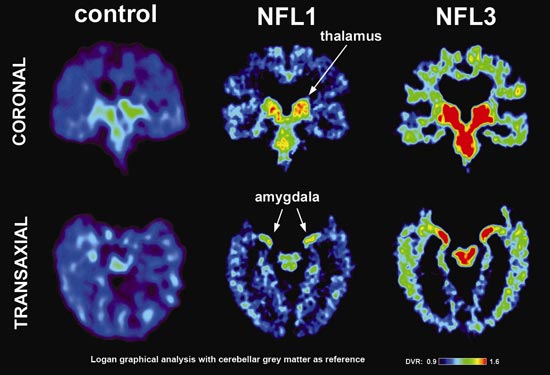
Elevated levels of FDDNP were observed in in the brains of 5 former NFL players relative to those of 5 control participants. The players were referred for testing because of symptoms of mild cognitive impairment. These five participants in the study (out of 19 potential volunteers) had a mean age of 59 yrs; MMSE scores of 28 vs. 30 in controls; and higher depression scores than controls, but no clinically significant depression in most.
The authors admit this is a very preliminary study that needs to be replicated and validated in larger samples. Yet they are optimistic they have a discovered a means of imaging CTE pathology in the brains of living patients.
But I would like to cast some doubt on this notion for two reasons:
(1) FDDNP is supposedly a tracer for both tau and amyloid beta (which forms plaques in Alzheimer's and other dementia), but some experts think it's neither. Studies have shown that it binds to a variety of misfolded proteins. For example, it selectively labels prion plaques in Creutzfeldt–Jakob disease (Bresjanac et al., 2003).
At any rate, FDDNP does not appear to be specific for CTE pathology.
(2) The distribution of FDDNP binding in the brains of the retired players does not appear to be the same as CTE tau pathology that has been observed at autopsy (McKee et al., 2012). Last month the Boston University group found evidence of CTE in the brains of in 68 male subjects, most of whom played contact sports. McKee et al. identified a series of four stages of CTE, with progressive worsening of clinical symptoms and neuropathology.
My guess is that the participants in the Small et al. study, if they do indeed have CTE, might be at stage I or perhaps stage II, but this is hard to tell given the very limited clinical and cognitive data on these patients (they had "a history of cognitive or mood symptoms").
The FDDNP results showed elevated signals in the players in a number of subcortical regions (including caudate, putamen, thalamus, subthalamus, midbrain, and amygdala), but nowhere in the cerebral cortex. On the other hand, stage I CTE tau pathology in post mortem brains is found in limited discrete locations: mild pathology in the cerebral cortex and minimal pathology in the amygdala and hippocampus (McKee et al., 2012). In stage II, pathology within the cortex spreads, and pathology in the medial temporal lobe (amygdala and hippocampus) is still mild.
Maybe there's a bias for subcortical labelling with FDDNP PET, versus cortical pTDP-43 immunoreactivity in post-mortem tissue. 3 That's possible, but Small and colleagues have previously reported extensive neocortical FDDND signal in mild cognitive impairment (Small et al., 2012).
I'm not an expert, and I could certainly be wrong about all of this. Development of in vivo imaging probes for neurodegenerative diseases is a very active area of research. The new hyperphosphorylated-tau radioligand [F-18]-T807 could be more specific for imaging tauopathies (Chien et al., 2012). Scientific developments like these will be especially crucial once any type of treatment for CTE comes along.
Footnotes
1 More background information, taken from my earlier post on Blast Wave Injury and Chronic Traumatic Encephalopathy: What's the Connection?
3 Noah Gray raised this point.
References
Bresjanac M, Smid LM, Vovko TD, Petric A, Barrio JR, Popovic M. (2003). Molecular-imaging probe 2-(1-[6-[(2-fluoroethyl)(methyl)amino]-2-naphthyl]ethylidene) malononitrile labels prion plaques in vitro. J Neurosci. 23(22):8029-33.
Chien DT, Bahri S, Szardenings AK, Walsh JC, Mu F, Su MY, Shankle WR, Elizarov A, Kolb HC. Early Clinical PET Imaging Results with the Novel PHF-Tau Radioligand[F-18]-T807. J Alzheimers Dis. 2012 Dec 12. [Epub ahead of print]
McKee, A., Stein, T., Nowinski, C., Stern, R., Daneshvar, D., Alvarez, V., Lee, H., Hall, G., Wojtowicz, S., Baugh, C., Riley, D., Kubilus, C., Cormier, K., Jacobs, M., Martin, B., Abraham, C., Ikezu, T., Reichard, R., Wolozin, B., Budson, A., Goldstein, L., Kowall, N., & Cantu, R. (2012). The spectrum of disease in chronic traumatic encephalopathy Brain DOI: 10.1093/brain/aws307
Gary W. Small, Vladimir Kepe, Prabha Siddarth, Linda M. Ercoli, David A. Merrill, Natacha Donoghue, Susan Y. Bookheimer, Jacqueline Martinez, Bennet Omalu, Julian Bailes, Jorge R. Barrio (2013). PET Scanning of Brain Tau in Retired National Football League Players: Preliminary Findings. Am J Geriatr Psychiatry, 21
- Fda Says No To Marketing Fddnp For Cte
The U.S. Food and Drug Administration recently admonished TauMark™, a brain diagnostics company, for advertising brain scans that can diagnose chronic traumatic encephalopathy (CTE), Alzheimer's disease, and other types of dementia. The Los Angeles...
- Blast Wave Injury And Chronic Traumatic Encephalopathy: What's The Connection?
Fig. 3 (Goldstein et al., 2012). Single-blast exposure induces CTE-like neuropathology in wild-type C57BL/6 mice. In a tour de force, a group of 35 Boston-area scientists1 (Goldstein et al., 2012) developed a mouse model of blast-related neurotrauma...
- Neuropsychology Abstract Of The Day: Olfaction And Dementia
Hubbard PS, Esiri MM, Reading M, McShane R, & Nagy Z. Alpha-synuclein pathology in the olfactory pathways of dementia patients. Journal of Anatomy. 2007 Jun 6; [Epub ahead of print] Division of Neuroscience, The Medical School, University of Birmingham,...
- Early Diagnosis Of Alzheimer Disease And Fddnp
From today's New York Times: New Chemical Is Said to Provide Early Sign of Alzheimer’s Disease By REUTERS The New York Times Published: December 21, 2006 chem Originally uploaded by rissera. A chemical designed by doctors in Los Angeles...
- Concussion Study Among Athletes
Twelve retired sports players have pledged to donate their brains to Boston University’s Center for the Study of Traumatic Encephalopathy (at BU's School of Medicine) which is devoted to studying the long-term effects of concussions. The Center...
Neuroscience
Is CTE Detectable in Living NFL Players?

Chronic traumatic encephalopathy (CTE) is a progressive neurodegenerative disease seen most often in athletes with repeated concussions.1 The condition has drawn extensive media attention due to the number of cases reported among retired NFL players. The disease can only be diagnosed at autopsy, because the brain tissue has to be stained for characteristic protein abnormalities which cannot be visualized in a living human.
Until now, that is, according to a new study by Gary Small and colleagues at UCLA (Small et al., 2013). Positron emission tomography (PET) and the molecular imaging probe FDDNP 2 were use to visualize levels of the protein tau, which forms neurofibrillary tangles in Alzheimer's disease and other tauopathies. Or what is presumed to be tau.
Elevated levels of FDDNP were observed in in the brains of 5 former NFL players relative to those of 5 control participants. The players were referred for testing because of symptoms of mild cognitive impairment. These five participants in the study (out of 19 potential volunteers) had a mean age of 59 yrs; MMSE scores of 28 vs. 30 in controls; and higher depression scores than controls, but no clinically significant depression in most.
The authors admit this is a very preliminary study that needs to be replicated and validated in larger samples. Yet they are optimistic they have a discovered a means of imaging CTE pathology in the brains of living patients.
But I would like to cast some doubt on this notion for two reasons:
(1) FDDNP is supposedly a tracer for both tau and amyloid beta (which forms plaques in Alzheimer's and other dementia), but some experts think it's neither. Studies have shown that it binds to a variety of misfolded proteins. For example, it selectively labels prion plaques in Creutzfeldt–Jakob disease (Bresjanac et al., 2003).
At any rate, FDDNP does not appear to be specific for CTE pathology.
(2) The distribution of FDDNP binding in the brains of the retired players does not appear to be the same as CTE tau pathology that has been observed at autopsy (McKee et al., 2012). Last month the Boston University group found evidence of CTE in the brains of in 68 male subjects, most of whom played contact sports. McKee et al. identified a series of four stages of CTE, with progressive worsening of clinical symptoms and neuropathology.
My guess is that the participants in the Small et al. study, if they do indeed have CTE, might be at stage I or perhaps stage II, but this is hard to tell given the very limited clinical and cognitive data on these patients (they had "a history of cognitive or mood symptoms").
The FDDNP results showed elevated signals in the players in a number of subcortical regions (including caudate, putamen, thalamus, subthalamus, midbrain, and amygdala), but nowhere in the cerebral cortex. On the other hand, stage I CTE tau pathology in post mortem brains is found in limited discrete locations: mild pathology in the cerebral cortex and minimal pathology in the amygdala and hippocampus (McKee et al., 2012). In stage II, pathology within the cortex spreads, and pathology in the medial temporal lobe (amygdala and hippocampus) is still mild.
Maybe there's a bias for subcortical labelling with FDDNP PET, versus cortical pTDP-43 immunoreactivity in post-mortem tissue. 3 That's possible, but Small and colleagues have previously reported extensive neocortical FDDND signal in mild cognitive impairment (Small et al., 2012).
I'm not an expert, and I could certainly be wrong about all of this. Development of in vivo imaging probes for neurodegenerative diseases is a very active area of research. The new hyperphosphorylated-tau radioligand [F-18]-T807 could be more specific for imaging tauopathies (Chien et al., 2012). Scientific developments like these will be especially crucial once any type of treatment for CTE comes along.
- Original link to study via Deadspin and Nature editor Noah Gray.
Footnotes
1 More background information, taken from my earlier post on Blast Wave Injury and Chronic Traumatic Encephalopathy: What's the Connection?
A defining pathological feature is tauopathy - abnormal accumulations of the tau protein seen in other dementias (e.g., Alzheimer's disease). Aggregations of hyperphosphorylated tau into neurofibrillary tangles (NFTs) are a defining feature, as in frontotemporal lobar degeneration and amyotrophic lateral sclerosis -- yet CTE is distinct from both of these (McKee et al., 2009). CTE results in cognitive and behavioral changes including memory impairments, poor impulse control, alterations in mood, suicidal behavior, disorientation, and ultimately dementia.2 Co-authors Small and Barrio are among the inventors of the FDDNP approach. They have received royalties and may receive royalties on future sales.
3 Noah Gray raised this point.
References
Bresjanac M, Smid LM, Vovko TD, Petric A, Barrio JR, Popovic M. (2003). Molecular-imaging probe 2-(1-[6-[(2-fluoroethyl)(methyl)amino]-2-naphthyl]ethylidene) malononitrile labels prion plaques in vitro. J Neurosci. 23(22):8029-33.
Chien DT, Bahri S, Szardenings AK, Walsh JC, Mu F, Su MY, Shankle WR, Elizarov A, Kolb HC. Early Clinical PET Imaging Results with the Novel PHF-Tau Radioligand[F-18]-T807. J Alzheimers Dis. 2012 Dec 12. [Epub ahead of print]
McKee, A., Stein, T., Nowinski, C., Stern, R., Daneshvar, D., Alvarez, V., Lee, H., Hall, G., Wojtowicz, S., Baugh, C., Riley, D., Kubilus, C., Cormier, K., Jacobs, M., Martin, B., Abraham, C., Ikezu, T., Reichard, R., Wolozin, B., Budson, A., Goldstein, L., Kowall, N., & Cantu, R. (2012). The spectrum of disease in chronic traumatic encephalopathy Brain DOI: 10.1093/brain/aws307
Gary W. Small, Vladimir Kepe, Prabha Siddarth, Linda M. Ercoli, David A. Merrill, Natacha Donoghue, Susan Y. Bookheimer, Jacqueline Martinez, Bennet Omalu, Julian Bailes, Jorge R. Barrio (2013). PET Scanning of Brain Tau in Retired National Football League Players: Preliminary Findings. Am J Geriatr Psychiatry, 21
- Fda Says No To Marketing Fddnp For Cte
The U.S. Food and Drug Administration recently admonished TauMark™, a brain diagnostics company, for advertising brain scans that can diagnose chronic traumatic encephalopathy (CTE), Alzheimer's disease, and other types of dementia. The Los Angeles...
- Blast Wave Injury And Chronic Traumatic Encephalopathy: What's The Connection?
Fig. 3 (Goldstein et al., 2012). Single-blast exposure induces CTE-like neuropathology in wild-type C57BL/6 mice. In a tour de force, a group of 35 Boston-area scientists1 (Goldstein et al., 2012) developed a mouse model of blast-related neurotrauma...
- Neuropsychology Abstract Of The Day: Olfaction And Dementia
Hubbard PS, Esiri MM, Reading M, McShane R, & Nagy Z. Alpha-synuclein pathology in the olfactory pathways of dementia patients. Journal of Anatomy. 2007 Jun 6; [Epub ahead of print] Division of Neuroscience, The Medical School, University of Birmingham,...
- Early Diagnosis Of Alzheimer Disease And Fddnp
From today's New York Times: New Chemical Is Said to Provide Early Sign of Alzheimer’s Disease By REUTERS The New York Times Published: December 21, 2006 chem Originally uploaded by rissera. A chemical designed by doctors in Los Angeles...
- Concussion Study Among Athletes
Twelve retired sports players have pledged to donate their brains to Boston University’s Center for the Study of Traumatic Encephalopathy (at BU's School of Medicine) which is devoted to studying the long-term effects of concussions. The Center...
