Neuroscience
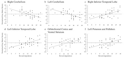
Or I would be if I had greater gray matter density in my left and right inferior temporal lobes, my orbitofrontal cortex and ventral striatum (collapsed into one big region of interest), and my left putamen and pallidum. Plus lower gray matter density in my left and right cerebellum (Lebreton et al., 2009).

To be fair to the authors, they never used the expression "people person" in their paper. That was the BBC1, among other press outlets:
Voxel-based morphometry was used to create probabilistic maps of gray matter for each individual. Associations between RD and brain structure were corrected for total gray matter volume and tested by fitting a multiple linear regression model at each voxel, followed by permutation testing. Out of the blue (with no previous or subsequent mention), the authors also decided to adjust for novelty seeking and harm avoidance. Lebreton et al. found the predicted correlation of RD and GMD in the orbitofrontal cortex (OFC) and the ventral striatum. They didn't have much to say about why the inferior temporal gyrus2 and cerebellum showed positive/negative correlations with RD, though. In the BBC article,

Footnotes
1 Found via @vaughanbell.
2 They called this region the temporal poles, but it looked more like ITG to me.
Reference

Lebreton, M., Barnes, A., Miettunen, J., Peltonen, L., Ridler, K., Veijola, J., Tanskanen, P., Suckling, J., Jarvelin, M., Jones, P., Isohanni, M., Bullmore, E., & Murray, G. (2009). The brain structural disposition to social interaction. European Journal of Neuroscience DOI: 10.1111/j.1460-9568.2009.06782.x.
- Does "internet Addiction" Really Shrink Your Brain?
Internet addiction is a murky and controversial disorder that is the subject of intense debate over whether it should be included in the new DSM-V. Here are the proposed diagnostic criteria as developed by Dr. Kimberly Young: Do you feel preoccupied...
- Jumping Into "the Fray" On Cerebral Asymmetry And Sexual Orientation
Fig. S1 (Savic & Lindström, 2008). Part of the left cerebral hemisphere VOI in a male heterosexual subject. Subject’s right side is to the right in the image. At The Fray, a reader discussion forum at Slate Magazine, a knowledgeable commenter...
- Employment Opportunity As A Professional Fmri Subject
Apply now! Or at least, that's the implication of this BBC story about the latest neuroimaging paper (Fliessbach et al., 2007) in Science: Men motivated by 'superior wage' [NOTE: so I guess women aren't, eh? we don't know, since they...
- Neuropsychology Abstract Of The Day: A Leisurely Curiousity
Training-induced neural plasticity in golf novices Journal of Neuroscience. 2011 Aug 31; 31(35): 12444-8. Authors: Bezzola L, Mérillat S, Gaser C, Jäncke L Abstract Previous neuroimaging studies in the field of motor learning have shown that learning...
- Neuropsychology Abstract Of The Day: Mild Cognitive Impairment (mci)
Karas G, Sluimer J, Goekoop R, van der Flier W, Rombouts S, Vrenken H, Scheltens P, Fox N, & Barkhof F. Amnestic Mild Cognitive Impairment: Structural MR Imaging Findings Predictive of Conversion to Alzheimer Disease. American Journal of Neuroradiology....
Neuroscience
I'm a People Person!
Or I would be if I had greater gray matter density in my left and right inferior temporal lobes, my orbitofrontal cortex and ventral striatum (collapsed into one big region of interest), and my left putamen and pallidum. Plus lower gray matter density in my left and right cerebellum (Lebreton et al., 2009).

click figure for larger view
Fig. 1 (from Lebreton et al., 2009). Regions in which gray matter density (GMD) is associated with reward dependence. Mean gray matter density was extracted from each of the clusters that we identified using linear regression, transformed into Z-scores, and plotted versus RD. The lines represent the best fit for the associations when adjusted for total gray matter volume.To be fair to the authors, they never used the expression "people person" in their paper. That was the BBC1, among other press outlets:
'People-person' brain area foundScientists say they have located the brain areas that may determine how sociable a person is. Warm, sentimental people tend to have more brain tissue in the outer strip of the brain just above the eyes and in a structure deep in the brain's centre. These are the same zones that allow us to enjoy chocolate and sex, the Cambridge University experts report in the European Journal of Neuroscience.. . .The men who scored higher on questionnaire-based ratings of emotional warmth and sociability had more grey matter in two brain areas - the orbitofrontal cortex and ventral striatum.As we can see in Figure 1, however, the gray matter volume in other brain areas (as measured using voxel-based morphometry) was also correlated with social reward dependence, defined as
a stable pattern of attitudes and behaviour hypothesized to represent a favourable disposition towards social relationships and attachment as a personality dimension.Social reward dependence (RD) was measured using
the RD scale of Cloninger’s temperament and character inventory (TCI), a questionnaire that maps independent temperament traits onto putatively independent neurobiological systems (Cloninger, 1987, 1994). The RD subscale measures the tendency of subjects to be sensitive to a socially defined reward: a high score indicates a high disposition to social relationships and attachment; and a low score indicates a tendency to insensitivity and aloofness.The participants were drawn from a large cohort of Finnish people born in 1966:
Data on biological, socioeconomic and health conditions, living habits, and family characteristics of cohort members were collected prospectively from pregnancy. The present study is based on 10,934 individuals living in Finland at the age of 16 years who did not forbid the use of their data...Of that gigantic cohort, the authors restricted subject selection to 4,349 people (1,974 males) who had completed the TCI questionnaire. Of the 1,531 living in the city of Oulu, 187 subjects were randomly selected and invited to participate, and 104 (62 men) agreed to have an MRI. The authors excluded female participants from the present study because they're more emotional and different from men [basically]. And it turned out that TCI data were not available from 21 of the 62 men after all, so the final sample was comprised of 41 male volunteers.
Voxel-based morphometry was used to create probabilistic maps of gray matter for each individual. Associations between RD and brain structure were corrected for total gray matter volume and tested by fitting a multiple linear regression model at each voxel, followed by permutation testing. Out of the blue (with no previous or subsequent mention), the authors also decided to adjust for novelty seeking and harm avoidance. Lebreton et al. found the predicted correlation of RD and GMD in the orbitofrontal cortex (OFC) and the ventral striatum. They didn't have much to say about why the inferior temporal gyrus2 and cerebellum showed positive/negative correlations with RD, though. In the BBC article,
Will we now see neurotraining programs designed to increase the size of your OFC and ventral striatum?Professor Simon Baron Cohen, of the Autism Research Centre in Cambridge, said: "This is an important study in showing that the degree to which we find socializing rewarding is correlated with differences in brain structure.
"It reminds us that for some people, socializing is an intrinsic reward, just like chocolate or cannabis. And that what you find rewarding depends on differences in the brain.

Footnotes
1 Found via @vaughanbell.
2 They called this region the temporal poles, but it looked more like ITG to me.
Reference

Lebreton, M., Barnes, A., Miettunen, J., Peltonen, L., Ridler, K., Veijola, J., Tanskanen, P., Suckling, J., Jarvelin, M., Jones, P., Isohanni, M., Bullmore, E., & Murray, G. (2009). The brain structural disposition to social interaction. European Journal of Neuroscience DOI: 10.1111/j.1460-9568.2009.06782.x.
- Does "internet Addiction" Really Shrink Your Brain?
Internet addiction is a murky and controversial disorder that is the subject of intense debate over whether it should be included in the new DSM-V. Here are the proposed diagnostic criteria as developed by Dr. Kimberly Young: Do you feel preoccupied...
- Jumping Into "the Fray" On Cerebral Asymmetry And Sexual Orientation
Fig. S1 (Savic & Lindström, 2008). Part of the left cerebral hemisphere VOI in a male heterosexual subject. Subject’s right side is to the right in the image. At The Fray, a reader discussion forum at Slate Magazine, a knowledgeable commenter...
- Employment Opportunity As A Professional Fmri Subject
Apply now! Or at least, that's the implication of this BBC story about the latest neuroimaging paper (Fliessbach et al., 2007) in Science: Men motivated by 'superior wage' [NOTE: so I guess women aren't, eh? we don't know, since they...
- Neuropsychology Abstract Of The Day: A Leisurely Curiousity
Training-induced neural plasticity in golf novices Journal of Neuroscience. 2011 Aug 31; 31(35): 12444-8. Authors: Bezzola L, Mérillat S, Gaser C, Jäncke L Abstract Previous neuroimaging studies in the field of motor learning have shown that learning...
- Neuropsychology Abstract Of The Day: Mild Cognitive Impairment (mci)
Karas G, Sluimer J, Goekoop R, van der Flier W, Rombouts S, Vrenken H, Scheltens P, Fox N, & Barkhof F. Amnestic Mild Cognitive Impairment: Structural MR Imaging Findings Predictive of Conversion to Alzheimer Disease. American Journal of Neuroradiology....
