Neuroscience
 i miss you
As Björk sings about her temporal conundrum, let's continue our time tunnel from the other day. Two recent neuroimaging papers have related the past to the future, as far as the brain is concerned. In the first study (Szpunar et al., 2007), participants were presented with a bunch of cue words (e.g., birthday, getting lost, etc) in the scanner and were told to orient their attention to the cue event. Then they had to either think about the event in relation to their future (SF), in relation to their past (SR), or in relation to a familiar individual (CI). The latter was a snappy and fun control condition: imagine Bill Clinton in that situation! OK, so comparisons were made between self-remember (SR), self-future (SF), and Clinton-imagine (CI). A set of regions (A-D), illustrated below in the left side of the figure, were more active for the future than the past. Another set of regions, illustrated on the right, had statistically equivalent activation when participants remembered the past and imagined the future.

from Szpunar et al. (2007)
The regions that were more active in imaging one's own future (not Bill's or Hillary's) included left lateral premotor cortex, our friend the precuneus in the left hemisphere, and the right cerebellum. [Note that left frontal cortex and right cerebellum are interconnected (Leiner et al., 1989; Niimura et al., 1999; Jansen et al., 2005]. The authors speculate that activation in a circuit that represents imagined motor movements is greater for novel movements that haven't occurred yet than for remembered motor sequences... hmmm.
Then there was a set of regions equally active when placing oneself in the past and in the future. These included elements of the dreaded default network (medial prefrontal, posterior cingulate). In fact, for the medial prefrontal ROI in panel E above, there was "less deactivation" (actually, the hemodynamic response looks flat and at zero) for self-related conditions than for the Clinton condition. More interesting are the other 3 regions: the parahippocampal gyrus (part of the medial temporal lobe memory system and home to the parahippocampal place area), posterior cingulate, and occipital cortex. The authors link these areas to autobiographical memories and spatial navigation. And perhaps, to Endel Tulving's conception of autonoetic consciousness, the ability to mentally represent and become aware of subjective experiences in the past, present, and future.


Parallel cerebello-frontal connections in the human brain. (Different parts of the cerebellum are linked to different areas of the frontal lobe. Because these connections enable the frontal areas to send signals to the cerebellum and receive signals from it, the cerebellum can serve as an information-processing adjunct to all of these frontal areas. Information can be processed repeatedly in such cerebro-cerebellar loops during the waking hours of life.)
This article generated a lot of press coverage, and some of it was actually fairly decent.
References
Jansen A, Floel A, Van Randenborgh J, Konrad C, Rotte M, Forster AF, Deppe M, Knecht S. (2005). Crossed cerebro-cerebellar language dominance. Hum Brain Mapp. 24: 165-72.
Leiner HC, Leiner AL, Dow RS. (1989). Reappraising the cerebellum: what does the hindbrain contribute to the forebrain? Behav Neurosci.103: 998-1008.
Niimura K, Chugani DC, Muzik O, Chugani HT. (1999). Cerebellar reorganization following cortical injury in humans: effects of lesion size and age. Neurology 52: 792-7.
- My Amygdala Is Very Optimistic Today...
...and my rostral anterior cingulate cortex imagines a brighter tomorrow. I don't know what all those pigs are doing in the posterior half of the brain, however. Oink. Doesn't everyone love a forcefully-worded headline? Source of ‘optimism’...
- The Overuse Theory Of Alzheimer's Disease
"Past and Future Self" by Austin Houldsworth Exploring the parallels between an expanding/contracting universe and human development.Shown at the 2006 Darklight Festival (among other places). View the 1 min video at perpetual art machine. As part of...
- Present Tense
Lately, The Neurocritic (and recent neuronews) has been focused on the past and the future. But what about the present? What happens to the brain when we are truly focused on the present moment? Overlapping brain areas activated while remembering the...
- Fables Of The Reconstruction
A flurry of papers (well, OK, three) has been published recently on the relationship between how we remember the past and imagine the future. The third and final paper is another functional neuroimaging study (Addis et al., 2007). Before we begin, let’s...
- Daydreaming And Thought-sampling
OK, is there anything new in the daydreaming article in Science? Fig. 2. Graphs depict regions that exhibited a significant positive relation [with a propensity to daydream], r(14) > 0.50, P < .05 (A) Bilateral mPFC; (B) Bilateral precuneus and posterior...
Neuroscience
i miss you but i haven't met you yet
 i miss you
but i haven't met you yet
so special
but it hasn't happened yet
you are gorgeous
but i haven't met you yet
i remember
but it hasn't happened yet
so special
but it hasn't happened yet
you are gorgeous
but i haven't met you yet
i remember
but it hasn't happened yet
-- Björk, I Miss You
Remembering The Past, Envisioning The Future
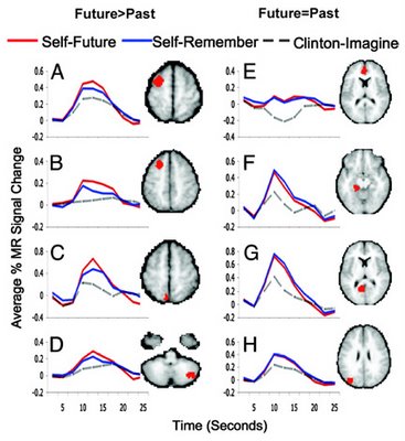
As Björk sings about her temporal conundrum, let's continue our time tunnel from the other day. Two recent neuroimaging papers have related the past to the future, as far as the brain is concerned. In the first study (Szpunar et al., 2007), participants were presented with a bunch of cue words (e.g., birthday, getting lost, etc) in the scanner and were told to orient their attention to the cue event. Then they had to either think about the event in relation to their future (SF), in relation to their past (SR), or in relation to a familiar individual (CI). The latter was a snappy and fun control condition: imagine Bill Clinton in that situation! OK, so comparisons were made between self-remember (SR), self-future (SF), and Clinton-imagine (CI). A set of regions (A-D), illustrated below in the left side of the figure, were more active for the future than the past. Another set of regions, illustrated on the right, had statistically equivalent activation when participants remembered the past and imagined the future.

from Szpunar et al. (2007)
| Future > Past | Future = Past |
| (A) L Middle Frontal | (E) Medial Frontal |
| (B) L Middle Frontal | (F) Parahippocampal Gyrus |
| (C) L Precuneus | (G) Posterior Cingulate |
| (D) R Cerebellum | (H) L |
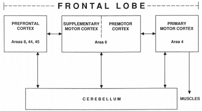
The regions that were more active in imaging one's own future (not Bill's or Hillary's) included left lateral premotor cortex, our friend the precuneus in the left hemisphere, and the right cerebellum. [Note that left frontal cortex and right cerebellum are interconnected (Leiner et al., 1989; Niimura et al., 1999; Jansen et al., 2005]. The authors speculate that activation in a circuit that represents imagined motor movements is greater for novel movements that haven't occurred yet than for remembered motor sequences... hmmm.
Then there was a set of regions equally active when placing oneself in the past and in the future. These included elements of the dreaded default network (medial prefrontal, posterior cingulate). In fact, for the medial prefrontal ROI in panel E above, there was "less deactivation" (actually, the hemodynamic response looks flat and at zero) for self-related conditions than for the Clinton condition. More interesting are the other 3 regions: the parahippocampal gyrus (part of the medial temporal lobe memory system and home to the parahippocampal place area), posterior cingulate, and occipital cortex. The authors link these areas to autobiographical memories and spatial navigation. And perhaps, to Endel Tulving's conception of autonoetic consciousness, the ability to mentally represent and become aware of subjective experiences in the past, present, and future.

Karl K. Szpunar, Jason M. Watson, and Kathleen B. McDermott. Neural substrates of envisioning the future. Proc. Natl. Acad. Sci. Published online before print January 3, 2007. OPEN ACCESS ARTICLE.From Behavioral Neuroscience 103: 998-1008, October 1989.
The ability to envision specific future episodes is a ubiquitous mental phenomenon that has seldom been discussed in the neuroscience literature. In this study, subjects underwent functional MRI while using event cues (e.g., Birthday) as a guide to vividly envision a personal future event, remember a personal memory, or imagine an event involving a familiar individual. Two basic patterns of data emerged. One set of regions (e.g., within left lateral premotor cortex; left precuneus; right posterior cerebellum) was more active while envisioning the future than while recollecting the past (and more active in both of these conditions than in the task involving imagining another person). These regions appear similar to those emerging from the literature on imagined (simulated) bodily movements. A second set of regions (e.g., bilateral posterior cingulate; bilateral parahippocampal gyrus; left occipital cortex) demonstrated indistinguishable activity during the future and past tasks (but greater activity in both tasks than the imagery control task); similar regions have been shown to be important for remembering previously encountered visual-spatial contexts. Hence, differences between the future and past tasks are attributed to differences in the demands placed on regions that underlie motor imagery of bodily movements, and similarities in activity for these two tasks are attributed to the reactivation of previously experienced visual-spatial contexts. That is, subjects appear to place their future scenarios in well known visual-spatial contexts. Our results offer insight into the fundamental and little-studied capacity of vivid mental projection of oneself in the future.

Parallel cerebello-frontal connections in the human brain. (Different parts of the cerebellum are linked to different areas of the frontal lobe. Because these connections enable the frontal areas to send signals to the cerebellum and receive signals from it, the cerebellum can serve as an information-processing adjunct to all of these frontal areas. Information can be processed repeatedly in such cerebro-cerebellar loops during the waking hours of life.)
This article generated a lot of press coverage, and some of it was actually fairly decent.
Scan shows how brains plot futureBrain Uses Past to Peer Into Future
The Washington University team say that specific areas of the brain are active when thinking about upcoming events.
. . .
The researchers placed 21 volunteers into the MRI machine, then contrasted the scan results when they were asked to imagine vividly future events and recollect past memories.
The resulting images showed clear differences between a birthday already experienced, and a birthday yet to come.
In particular, when looking ahead, three particular areas of the brain were activated - the left lateral premotor cortex, the left precuneus and the right posterior cerebellum.
These brain areas are already known to be involved in the imagining of body movements, suggesting that when the human brain is thinking about the future, it does so in terms of distinct movements and actions that will happen at that point.
References
Jansen A, Floel A, Van Randenborgh J, Konrad C, Rotte M, Forster AF, Deppe M, Knecht S. (2005). Crossed cerebro-cerebellar language dominance. Hum Brain Mapp. 24: 165-72.
Leiner HC, Leiner AL, Dow RS. (1989). Reappraising the cerebellum: what does the hindbrain contribute to the forebrain? Behav Neurosci.103: 998-1008.
Niimura K, Chugani DC, Muzik O, Chugani HT. (1999). Cerebellar reorganization following cortical injury in humans: effects of lesion size and age. Neurology 52: 792-7.
- My Amygdala Is Very Optimistic Today...
...and my rostral anterior cingulate cortex imagines a brighter tomorrow. I don't know what all those pigs are doing in the posterior half of the brain, however. Oink. Doesn't everyone love a forcefully-worded headline? Source of ‘optimism’...
- The Overuse Theory Of Alzheimer's Disease
"Past and Future Self" by Austin Houldsworth Exploring the parallels between an expanding/contracting universe and human development.Shown at the 2006 Darklight Festival (among other places). View the 1 min video at perpetual art machine. As part of...
- Present Tense
Lately, The Neurocritic (and recent neuronews) has been focused on the past and the future. But what about the present? What happens to the brain when we are truly focused on the present moment? Overlapping brain areas activated while remembering the...
- Fables Of The Reconstruction
A flurry of papers (well, OK, three) has been published recently on the relationship between how we remember the past and imagine the future. The third and final paper is another functional neuroimaging study (Addis et al., 2007). Before we begin, let’s...
- Daydreaming And Thought-sampling
OK, is there anything new in the daydreaming article in Science? Fig. 2. Graphs depict regions that exhibited a significant positive relation [with a propensity to daydream], r(14) > 0.50, P < .05 (A) Bilateral mPFC; (B) Bilateral precuneus and posterior...
