Neuroscience
 Attention (everyone knows what that1 is) is often described as having "bottom-up" and "top-down" components (referring to the direction of information flow from sensory to high-order cortical regions, and vice versa).
ThinkGeek (bottoms up!)
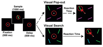
In a recent study appearing in Science, Buschman and Miller (2007) recorded simultaneously from a number of neurons in both frontal and parietal cortices to examine the onset and location of responses to the two types of attention, as summarized below.

from Buschman & Miller (2007)
Buschman TJ, Miller EK (2007). Top-Down Versus Bottom-Up Control of Attention in the Prefrontal and Posterior Parietal Cortices. Science 315: 1860-1862.
Attention can be focused volitionally by "top-down" signals derived from task demands and automatically by "bottom-up" signals from salient stimuli. The frontal and parietal cortices are involved, but their neural activity has not been directly compared. Therefore, we recorded from them simultaneously in monkeys. Prefrontal neurons reflected the target location first during top-down attention, whereas parietal neurons signaled it earlier during bottom-up attention. Synchrony between frontal and parietal areas was stronger in lower frequencies during top-down attention and in higher frequencies during bottom-up attention. This result indicates that top-down and bottom-up signals arise from the frontal and sensory cortex, respectively, and different modes of attention may emphasize synchrony at different frequencies.
So we see what was expected: top-down attention is top-down, and bottom-up attention is bottom-up. Have we learned anything we don't already know? What's novel about this study? Well, the technical feat of simultaneously recording from multiple single neurons in 3 different cortical regions is impressive, as are the detailed statistical analyses described in the 27 page supplement.
In the press, the authors stretched their point a bit to state the study's relevance to ADD:
Footnote
- Judging What Came First
You can’t possibly process everything that’s going on around you. Instead you’re armed with an attentional spotlight that selects areas and objects of interest for preferential processing. An anomalous consequence of this, is that we judge objects...
- The Brain Can't Ignore Angry Voices
We're highly tuned to emotional signals. So whereas most of the information bombarding our brain is filtered, emotion-related signals seem to strike home regardless. Take fearful faces - research has shown these trigger the same activity in a fear-sensitive...
- Mmmm... Donuts... The Sequel
% Simpson Tide, Act one. Homer is standing in chains before % a court composed completely of immense, talking doughnuts % of various shapes, colors, and flavors. The judge, % assumably, is a white doughnut seated at a table across % from him. White Doughnut:...
- Ready For Your Close-up, Mr. Chocolate Iced Kreme Filled
Your brain on Krispy Kremes CHICAGO--What makes you suddenly dart into the bakery when you spy chocolate- frosted donuts in the window, though you certainly hadn't planned on indulging? As you lick the frosting off your fingers, don't blame a...
- Neuropsychology Abstract Of The Day: Fmri Study Of Digit Symbol Task
Usui N, Haji T, Maruyama M, Katsuyama N, Uchida S, Hozawa A, Omori K, Tsuji I, Kawashima R, & Taira M. (2009). Cortical areas related to performance of WAIS Digit Symbol Test: a functional imaging study. Neurosci Lett. Jul 21. Many neuropsychological...
Neuroscience
Bottoms Up
 Attention (everyone knows what that1 is) is often described as having "bottom-up" and "top-down" components (referring to the direction of information flow from sensory to high-order cortical regions, and vice versa).
ThinkGeek (bottoms up!)
In a recent study appearing in Science, Buschman and Miller (2007) recorded simultaneously from a number of neurons in both frontal and parietal cortices to examine the onset and location of responses to the two types of attention, as summarized below.
Attention and Information FlowThe authors defined bottom-up attention as pop-out (or visual salience, an "automatic" process) and top-down attention as visual search (a "controlled" process). Thus, the flow of information was initially defined by task, not by physiology (Treisman & Gelade, 1980; pdf)
Cortical neurons modulate their activity with shifts in attention, but the source and flow of attention signals are unclear. Buschman et al. (p. 1860) used 50 electrodes to record simultaneously the activity from three cortical regions thought to be critical for attention. Bottom-up shifts of attention were first reflected in the parietal cortex, whereas top-down shifts of attention were reflected first in the frontal cortex. Thus, external control of visual attention originates in parietal cortex, but internal control of visual attention is directed from the frontal cortex.

from Buschman & Miller (2007)
Buschman TJ, Miller EK (2007). Top-Down Versus Bottom-Up Control of Attention in the Prefrontal and Posterior Parietal Cortices. Science 315: 1860-1862.
Attention can be focused volitionally by "top-down" signals derived from task demands and automatically by "bottom-up" signals from salient stimuli. The frontal and parietal cortices are involved, but their neural activity has not been directly compared. Therefore, we recorded from them simultaneously in monkeys. Prefrontal neurons reflected the target location first during top-down attention, whereas parietal neurons signaled it earlier during bottom-up attention. Synchrony between frontal and parietal areas was stronger in lower frequencies during top-down attention and in higher frequencies during bottom-up attention. This result indicates that top-down and bottom-up signals arise from the frontal and sensory cortex, respectively, and different modes of attention may emphasize synchrony at different frequencies.
So we see what was expected: top-down attention is top-down, and bottom-up attention is bottom-up. Have we learned anything we don't already know? What's novel about this study? Well, the technical feat of simultaneously recording from multiple single neurons in 3 different cortical regions is impressive, as are the detailed statistical analyses described in the 27 page supplement.
In the press, the authors stretched their point a bit to state the study's relevance to ADD:
Neuroscientists find different brain regions fuel attentionIn addition to charting the activity of single neurons, the authors recorded local field potentials (e.g., see Kreiman et al., 2006 among many others) and determined the frequency bands that showed coherence (neural synchrony) between frontal and parietal regions, observing
. . .
ADD involves being overly sensitive to the automatic attention-grabbers and less able to willfully sustain attention. "Our work suggests that we should target different parts of the brain to try to fix different types of attention deficits," Miller said.
"The downside of most psychiatic drugs is they are too broad," he continued. "It's like hitting the problem with a sledgehammer; you get the benefits but also many unintended consequences. Our work suggests that we may one day be able to figure out what is the exact problem with each individual and specifically target those shortcomings. And that is the ultimate goal in psychiatric intervention."
a greater increase in middle-frequency (22 to 34 Hz) coherence between LIP and frontal cortex during top-down search than during bottom-up pop-out. By contrast, the increase in upper-frequency (35 to 55 Hz) coherence was greater during pop-out than during search. Thus, bottom-up and top-down attention may rely on different frequency bands of coherence between the frontal and parietal cortex.What about EEG studies in humans? Intracranial recordings in epilepsy patients? What have these methodologies revealed about bottom-up and top-down attention? [And were any of those papers published in Science?] Stay tuned...
Footnote
1 Everyone knows what attention is. It is the taking possession by the mind in clear and vivid form, of one out of what seem several simultaneously possible objects or trains of thought...It implies withdrawal from some things in order to deal effectively with others.
-- William James
Principles of Psychology (1890)
- Judging What Came First
You can’t possibly process everything that’s going on around you. Instead you’re armed with an attentional spotlight that selects areas and objects of interest for preferential processing. An anomalous consequence of this, is that we judge objects...
- The Brain Can't Ignore Angry Voices
We're highly tuned to emotional signals. So whereas most of the information bombarding our brain is filtered, emotion-related signals seem to strike home regardless. Take fearful faces - research has shown these trigger the same activity in a fear-sensitive...
- Mmmm... Donuts... The Sequel
% Simpson Tide, Act one. Homer is standing in chains before % a court composed completely of immense, talking doughnuts % of various shapes, colors, and flavors. The judge, % assumably, is a white doughnut seated at a table across % from him. White Doughnut:...
- Ready For Your Close-up, Mr. Chocolate Iced Kreme Filled
Your brain on Krispy Kremes CHICAGO--What makes you suddenly dart into the bakery when you spy chocolate- frosted donuts in the window, though you certainly hadn't planned on indulging? As you lick the frosting off your fingers, don't blame a...
- Neuropsychology Abstract Of The Day: Fmri Study Of Digit Symbol Task
Usui N, Haji T, Maruyama M, Katsuyama N, Uchida S, Hozawa A, Omori K, Tsuji I, Kawashima R, & Taira M. (2009). Cortical areas related to performance of WAIS Digit Symbol Test: a functional imaging study. Neurosci Lett. Jul 21. Many neuropsychological...
