An "Endophenotype" For Sexual Orientation?
OR: The Neuroscience of Porn, according to The Frontal Cortex.
Wiktionary defines endophenotype as "any hereditary characteristic that is normally associated with some condition but is not a direct symptom of that condition."
Gottesman and Gould (2003) have this to say:
Endophenotypes, measurable components unseen by the unaided eye along the pathway between disease and distal genotype, have emerged as an important concept in the study of complex neuropsychiatric diseases. An endophenotype may be neurophysiological, biochemical, endocrinological, neuroanatomical, cognitive, or neuropsychological (including configured self-report data) in nature. Endophenotypes represent simpler clues to genetic underpinnings than the disease syndrome itself, promoting the view that psychiatric diagnoses can be decomposed or deconstructed, which can result in more straightforward—and successful—genetic analysis. However, to be most useful, endophenotypes for psychiatric disorders must meet certain criteria, including association with a candidate gene or gene region, heritability that is inferred from relative risk for the disorder in relatives, and disease association parameters. In addition to furthering genetic analysis, endophenotypes can clarify classification and diagnosis and foster the development of animal models. The authors discuss the etymology and strategy behind the use of endophenotypes in neuropsychiatric research and, more generally, in research on other diseases with complex genetics.However, for the study under discussion here, the "endophenotype" for sexual orientation is not diagnostic for a disorder of any sort, but merely for whether an individual (and his/her ventral premotor cortex) prefers to view pictures of the aroused genitals from one sex or the other. Do a Google seach for endophenotype, and the links are, in fact, mostly about disorders like schizophrenia, ADHD, autism, impulsivity, alcoholism, etc. Same for a PubMed search. Already we're dealing with a slightly loaded term, then, because the word endophenotype is associated with disorder.
Gottesman II, Gould TD (2003). The endophenotype concept in psychiatry: Etymology and strategic intentions. Am J Psychiatry 160:636-645.
But what about the pornography? Here it is,1 in NeuroImage:
Ponseti J, Bosinski HA, Wolff S, Peller M, Jansen O, Mehdorn HM, Buchel C, Siebner HR. A functional endophenotype for sexual orientation in humans. Neuroimage 2006 Sep 14; [Epub ahead of print]OK, let's look at the study's German participants. There were 12 straight men, 12 straight women (0 or 1 on the Kinsey scale, meaning exclusively heterosexual or predominantly heterosexual) and 15 gay men, 14 gay women (Kinsey 6, meaning exclusively homosexual or 7... what's a Kinsey 7?? maybe they meant 5 and 6?). Anyway, those in the middle were excluded (sorry, all you bisexual Kinsey 2-4's out there, left out again). Below is the Kinsey Scale:
Sexually arousing visual stimuli activate the human reward system and trigger sexual behavior. Here we performed event-related fMRI during visual processing of sexual core stimuli to pinpoint a neuronal correlate of sexual preference in humans. To dissociate gender of the stimulus from sexual preference, we studied male and female heterosexual and homosexual volunteers while they viewed sexual and nonsexual control stimuli. In contrast to previous work, we used core single-sex stimuli displaying male and female sexually aroused genitals. Since stimuli lacked any additional contextual information, they evoked no activity related to neuronal processing of faces, gestures or social interactions. Our prediction was that the sexual preference of the observer determines the neuronal responsiveness to pure male or female sexual stimuli in the human reward and motor system. Consistent with our prediction, the ventral striatum and the centromedian thalamus, showed a stronger neuronal response to preferred relative to non-preferred stimuli. Likewise, the ventral premotor cortex which is a key structure for imitative (mirror neurons) and tool-related (canonical neurons) actions showed a bilateral sexual preference-specific activation, suggesting that viewing sexually aroused genitals of the preferred sex triggers action representations of sexual behavior. The neuronal response of the ventral striatum, centromedian thalamus and ventral premotor cortex to preferred sexual stimuli was consistent across all groups. We propose that this invariant response pattern in core regions of the human reward and motor system represents a functional endophenotype for sexual orientation independent of the gender of the observer and gender of the stimulus.
| Rating | Description |
| 0 | Exclusively heterosexual |
| 1 | Predominantly heterosexual, only incidentally homosexual |
| 2 | Predominantly heterosexual, but more than incidentally homosexual |
| 3 | Equally heterosexual and homosexual |
| 4 | Predominantly homosexual, but more than incidentally heterosexual |
| 5 | Predominantly homosexual, only incidentally heterosexual |
| 6 | Exclusively homosexual |
So in the scanner, the participants viewed, in random order, 30 pictures of female genitals, 30 pics of male genitals, and 30 neutral stimuli. The task was to press a button when an oddball target stimulus appeared (a green square). Immediately after the scan, the subjects rated the visual stimuli on arousal and valence.

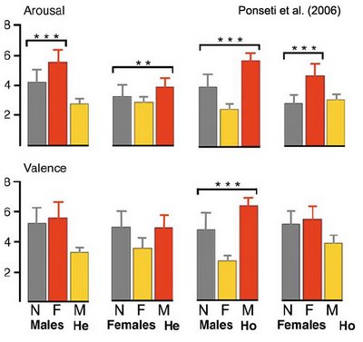
Arousal ratings conformed more or less to the predicted pattern. In Fig. 1 above, the post-hoc comparisons between nonsexual stimuli (gray bars) and preferred sexual stimuli (red bars) are significant for all groups (albeit to a smaller degree for the straight women). In general, ratings for preferred vs. nonpreferred sexual stimuli were more discrepant in men than in women (regardless of orientation).
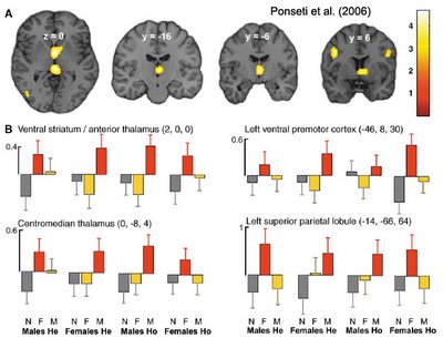
For the fMRI results, the authors defined 3 regions of interest, based on a priori hypotheses:
1. Centromedian Thalamus (and adjacent ventral striatum) - reward system [striatum, yes, but thalamus??]
2. Orbitofrontal Cortex - reward system
3. Ventral Premotor Cortex (PMv) - "motor representations of sexual behavior" [genital mirror neurons??]
Note that the amygdala was not among the ROIs, even though others have observed greater amygdala activity when viewing sexual stimuli compared to neutral stimuli (e.g., Hamann et al., 2004; see the Comments section of Sweat, Urine, and Sexual Orientation for a review of sex differences in brain activity when viewing porn clips). Come to think of it, the hypothalamus wasn't selected as an ROI, either. Hmmm, makes one wonder whether the ROIs were a posteriori...

Anyway, the red bars in the figure above (preferred sexual stimuli) depict greater activity than the yellow bars (nonpreferred sexual stimuli), which held for all four groups in thalamic regions, PMv, left superior parietal lobule [hey, what's that about??], but not orbitofrontal cortex.
Here's a thought. According to Wikipedia,
a mirror neuron is a neuron which fires both when an animal performs an action and when the animal observes the same action performed by another (especially conspecific) animal. Thus, the neuron "mirrors" the behavior of another animal, as though the observer were himself performing the action.The Neurocritic is as skeptical as anyone about the mirror neuron craze, but if the PMv activity in this experiment is really imitative in nature, or even "empathetic" (instead of motor imagery or motor preparation), then wouldn't same-sex genitals elicit greater activity than opposite-sex genitals, regardless of sexual orientation?
Never mind.
The foci of premotor activation closely corresponded to the site that was found to be activated by observation of hand or mouth movements (Buccino et al., 2001) and by the observation of action-related objects (Grezes et al., 2003).[NOTE: So what would happen if you showed pictures of sex toys or sexbots or or... never mind.]
The observation of sexually aroused genitals of the preferred sex may trigger neuronal activity of mirror neurons or canonical neurons in the PMv. Sexually aroused genitals of the preferred sex are targets of manipulative actions during mating. Thus, in analogy to tools [NOTE: !!], these stimuli can be conceptualized as action-related objects which may activate canonical neurons in the PMv. Beyond its significance as a tool-like target, a sexually aroused genital belongs to an agent and indicates the motivational and bodily state of the agent (i.e., the readiness to mate).
Ventral premotor mirror neurons may be involved in understanding the intention of the sexually preferred agent and activating a repertoire of volitional sexual actions.
[Hmm, seems like you could program a robot that way...]
But here's the bottom line:
We propose that this response reflect the activation of motor representations of manual or oral sexual behavior.
They really don't discuss the endophenotype idea much more, except to say,
We show a strong relation between the neuronal response and sexual orientation in distinct structures of the human reward and motor neuron system. ...the specific activity pattern induced by preferred sexual stimuli was remarkably invariant across heterosexual and homosexual males and females. In contrast to sexual preference, the sex of the stimulus and of the observer had no influence on the neuronal response to sexually aroused genitals. Therefore, we propose that the observed response pattern represents a functional endophenotype for sexual orientation in humans.
Moulier V, Mouras H, Pelegrini-Issac M, Glutron D, Rouxel R, Grandjean B, Bittoun J, Stoleru S. Neuroanatomical correlates of penile erection evoked by photographic stimuli in human males. Neuroimage 2006 Sep 6; [Epub ahead of print]
The objective of this study was to identify the cerebral correlates of the early phase, and of low to moderate levels, of penile tumescence using for the first time a volumetric measure of the penile response. We hypothesized that (i) regions whose response had been found correlated with circumferential penile responses in previous studies would be identified with volumetric plethysmography and (ii) that other brain regions, including the amygdalae, would be found using the more sensitive volumetric measurement. In ten healthy males, functional magnetic resonance imaging (fMRI) was used to study brain responses to sexually stimulating photographs and to various categories of control photographs. Both ratings of perceived erection and penile plethysmography demonstrated an erectile response to the presentation of sexually stimulating photographs. Regions where the BOLD signal was correlated with penile volumetric responses included the right medial prefrontal cortex, the right and left orbitofrontal cortices, the insulae, the paracentral lobules, the right ventral lateral thalamic nucleus, the right anterior cingulate cortex and regions involved in motor imagery and motor preparation (supplementary motor areas, left ventral premotor area). This study suggests that the development of low levels of penile tumescence in response to static sexual stimuli is controlled by a network of frontal, parietal, insular and cingulate cortical areas and that penile tumescence reciprocally induces activation in somatosensory regions of the brain.
1The paper did not provide any examples of the stimuli used in the experiment. The reviewers apparently missed out on the opportunity to request that such images appear in an Appendix. But here's the verbal description (Ponseti et al., 2006).First, sexual stimuli had to be specific to one or the other sex. Second, sexual stimuli should lack any further context information to avoid confounding brain activation related to neuronal processing of faces, gestures or social interactions. To meet these constraints we used photographs of naked male or female trunks displaying signs of genital arousal without showing the head or distal limbs. In addition we presented matched nonsexual photographs of the International Affective Picture System (IAPS) (Lang et al., 1997) as control stimuli. Male sexual stimuli showed an erected penis, female sexual stimuli showed details of the vulva (labia, clitoris, moist). The torso was sometimes visible together with the genitals, but the face or the limbs were never displayed on the photographs.
Buccino G, Binkofski F, Fink GR, Fadiga L, Fogassi L, Gallese V, Seitz RJ, Zilles K, Rizzolatti G, Freund HJ (2001). Action observation activates premotor and parietal areas in a somatotopic manner: an fMRI study. Eur. J. Neurosci. 13: 400-404.
Grezes J, Armony JL, Rowe J, Passingham RE (2003). Activations related to "mirror" and "canonical" neurones in the human brain: an fMRI study. NeuroImage 18: 928-937.
Hamann S, Herman RA, Nolan CL, Wallen K. (2004). Men and women differ in amygdala response to visual sexual stimuli. Nat Neurosci. 7: 411-6.
- How Messages Are Scent
The idea that humans release chemical signals – pheromones – that affect people around them is controversial. If pheromones do exist, however, two chemicals that might fulfil this role are the testosterone derivative 4,16-androstadien-3-one (AND),...
- Media Hsdd: "hyperactive Sexual Disorder Detection"
You might have seen the news stories with their facile headlines: Women with low libidos 'have different brains'Women with low libidos have a different mental response to intimate situations than those with a 'normal' sex drive, researchers...
- Spanner Or Sex Object?
Sex objects: Pictures shift men's view of women by Ian Sample, ChicagoMen are more likely to think of women as objects if they have looked at sexy pictures of females beforehand, psychologists said yesterday.Researchers used brain scans to show...
- Invisible Nudes Redux
No, the invisible nudes are not taking Redux [although visible nudes might be], rather the PNAS paper on sex, sexual orientation, and implicit attentional priming by naked pix has finally been posted online: Yi Jiang, Patricia Costello, Fang Fang, Miner...
- Sweat, Urine, And Sexual Orientation
Sexual Orientation May Affect Brain Response to Human Pheromones By Miranda Hitti WebMD Medical News Reviewed By Louise Chang, MD on Monday, May 08, 2006 May 8, 2006 -- Lesbian women and heterosexual women respond differently...
