Neuroscience
 Figure 3A (adapted from Komisaruk et al., 2011). Group-based composite view of the clitoral, vaginal, and cervical activation sites, all in the medial paracentral lobule, but regionally differentiated. We interpret this as due to the differential sensory innervation of these genital structures, i.e., clitoris: pudendal nerve, vagina: pelvic nerve,1 and cervix: hypogastric and vagus nerves.
"Femunulus" is a neologism for "female homuculus"
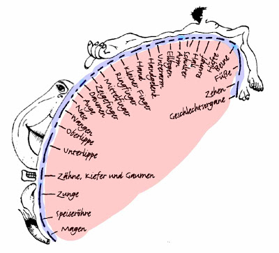
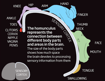
The neuroanatomical definition of homunculus is a "distorted" representation of the sensorimotor body map (and its respective parts) overlaid upon primary somatosensory and primary motor cortices. The figure below illustrates the sensory homunculus, where each body part is placed onto the region of cortex that represents it, and the size of the body part is proportional to its cortical representation (and sensitivity). It's rare to see the genitals represented at all. And if they are present, they are inevitably male genitals.
Homunculus image from Reinhard Blutner.
See the G-Rated [i.e., genital-less] flash explanation of homunculus.
A New Clitoral Homunculus?
To remedy this puritanical and androcentric situation, Swiss scientists at University Hospital in Zurich conducted a highly stimulating study in 15 healthy women to map the somatosensory representation of the clitoris (Michels et al., 2009).
Michels and colleagues began by reviewing the work of Wilder Penfield et al.:
However, that experiment involved electrical stimulation of the dorsal clitoral nerve, which was not sexually arousing. What if the stimulation occurred in a more naturalistic fashion?
Even newer clitoral, vaginal, cervical and nipple homunculi?
Now, Komisaruk et al., (2011) have expanded the somatosensory map of female sexual organs by having the participants engage in self-stimulation of the clitoris, vagina, cervix, and nipple while laying in a 3T scanner. For comparison, the investigators stimulated the thumb and the big toe. Should we be concerned about differential movement artifact (of the head, hand, arm, pelvis) in these varied stimulation conditions? We'll leave that question aside for the moment and examine the experimental protocol, which consisted of 30 sec of rest and 30 sec of the various stimulation modalities, each followed by 30 sec rest (in 5 min blocks):
 Figure 2 (adapted from Komisaruk et al., 2011). Three-axis (coronal, sagittal, and transaxial) views of the group-based responses to participant self-applied (clitoris, vagina, or cervix) stimulation ... The arrows indicate the sensory cortical regions activated by the various stimulated body regions. Clitoral, vaginal, and cervical self-stimulation activated the medial paracentral lobule. Note that the perineal (groin) region just lateral to the midline in the paracentral lobule was also activated... There was marked hand-related activation in the postcentral gyrus, and continuation of activation into the supplementary motor area immediately rostral to the sensory cortical responses... The secondary sensory cortex (SII; at the base of the homunculus) was activated under all the stimulus conditions...
To me, it looks like all the midline activations [including the toe activation, not shown here] bleed into motor areas including supplementary motor cortex, which is involved in both planning and inhibiting movement. We aren't given MNI coordinates to precisely locate the activations in 3D space, but my guess is that the clitoral, vaginal, and cervical regions are more overlapping than Figure 3A would lead you to believe. In fact, there is visible overlap between the vaginal and cervical regions, and the authors say this is due to unavoidable cross-stimulation in the cervical condition. Given the amount of motion artifact that likely occurred, a 4-5 mm difference in the foci of activation may not be entirely reliable in this particular study.3
The most surprising finding was that nipple stimulation largely overlapped with the genital regions:
Footnote
1 I'm not sure there is one "pelvic nerve" per se in the human female, as opposed to in rats.
2 You'll notice that Figure 3A better illustrates the bilateral nature of the activation, however [which could suggest mirror movement on the L side].
3 I'm not familiar enough with the human peripheral nervous system to say whether separate nerves innervate different female sexual regions with no overlap.
References
Kell CA, von Kriegstein K, Rösler A, Kleinschmidt A, Laufs H. (2005). The sensory cortical representation of the human penis: revisiting somatotopy in the male homunculus. J Neurosci. 25:5984-7.
Komisaruk, B., Wise, N., Frangos, E., Liu, W., Allen, K., & Brody, S. (2011). Women's Clitoris, Vagina, and Cervix Mapped on the Sensory Cortex: fMRI Evidence. The Journal of Sexual Medicine DOI: 10.1111/j.1743-6109.2011.02388.x
Michels, L., Mehnert, U., Boy, S., Schurch, B., & Kollias, S. (2009). The somatosensory representation of the human clitoris: An fMRI study NeuroImage.
 Body mapping on the brain - revised homunculus from New Scientist.
- Extras
Eye-catching studies that didn't make the final cut: A longitudinal study of children's text messaging and literacy development. First ever mapping of women's genitals as represented in the sensory cortex of the female brain. "Vaginal, clitoral,...
- Feeling Other People's Pain
When we watch someone else being pricked by a needle in their hand, the corticospinal motor neurons connected to that specific part of our own hand are inhibited, just as they would be if we’d been injected ourselves. It’s as though our brain has...
- More Exciting Than 'female Orgasm Captured In Series Of Brain Scans'
The intrinsic functional connectivity networks of human lateral frontal cortex! "The intrinsic functional connectivity networks of human lateral frontal cortex are displayed for a 4-mm seed region that is gradually moved along the cortical surface....
- A New Penile Homunculus?
Homunculus image from Reinhard Blutner. Leave it to those wacky writers at LiveScience.com to come up with a sure-to-click headline: Study: Circumcision Removes Most Sensitive Parts By Ker Than, LiveScience Staff Writer posted: 15 June 2007 12:53 pm...
- Alternative To Deep-brain Stimulation In Parkinson Disease?
From Reuters:Brain Surface Stimulation May Ease Parkinson's Mon Jan 3, 2005 06:22 PM GMT By Anne Harding NEW YORK (Reuters Health) - Electrical stimulation of regions deep in the brain has become fairly common in recent years for treating Parkinson's...
Neuroscience
A New Sexual Femunculus?
Figure 3A (adapted from Komisaruk et al., 2011). Group-based composite view of the clitoral, vaginal, and cervical activation sites, all in the medial paracentral lobule, but regionally differentiated. We interpret this as due to the differential sensory innervation of these genital structures, i.e., clitoris: pudendal nerve, vagina: pelvic nerve,1 and cervix: hypogastric and vagus nerves."Femunulus" is a neologism for "female homuculus"
The neuroanatomical definition of homunculus is a "distorted" representation of the sensorimotor body map (and its respective parts) overlaid upon primary somatosensory and primary motor cortices. The figure below illustrates the sensory homunculus, where each body part is placed onto the region of cortex that represents it, and the size of the body part is proportional to its cortical representation (and sensitivity). It's rare to see the genitals represented at all. And if they are present, they are inevitably male genitals.
Homunculus image from Reinhard Blutner.
See the G-Rated [i.e., genital-less] flash explanation of homunculus.
A New Clitoral Homunculus?
To remedy this puritanical and androcentric situation, Swiss scientists at University Hospital in Zurich conducted a highly stimulating study in 15 healthy women to map the somatosensory representation of the clitoris (Michels et al., 2009).
Michels and colleagues began by reviewing the work of Wilder Penfield et al.:
During the last 70 years the description of the sensory homunculus has been virtually a standard reference for various somatotopical studies (Penfield and Boldrey 1937; PDF). This map consists of a detailed description of the functional cortical representation of different body parts obtained via electrical stimulation during open brain surgery. In their findings they relied on reported sensations of different body parts after electrical stimulation of the cortex. Assessment of the exact location was generally difficult and sometimes led to conflicting results. The genital region was especially hard to assess due to difficulties with sense of shame.In contrast to electrical stimulation of the brain, modern mapping studies have used sensory stimulation to map the penis with fMRI (e.g., Kell et al., 2005). But as of 2009, there were no comparable fMRI studies of female genitalia. So how is such a study conducted, methodologically speaking? Electrical stimulation of the dorsal clitoral nerve was compared to electrical stimulation of the hallux (big toe). It was all very clinical, no sexual arousal involved. Here's the experimental protocol (Michels et al., 2009):
Prior to the imaging session, two self-attaching surface disc electrodes (1 × 1 cm) were placed bilaterally next to the clitoris of the subjects so that we were able to stimulate the fibers of the dorsal clitoral nerve. Before the start of the experiment, electrical test stimulation was performed to ensure that subjects could feel the stimulation directly at the clitoris. In addition, the strength of electrical stimulation was adjusted to a subject-specific level, i.e. that stimulation was neither felt [as] painful nor elicited – in case of clitoris stimulation – any sexual arousal. Functional imaging was performed in a block design with alternating rest and stimulation conditions, starting with a rest condition. ... In addition to the clitoris stimulation, we performed in eight of the recorded subjects a second experimental session, in which we applied electrical stimulation of the right hallux using the same type of electrodes, stimulation and scan paradigm.Their neuroimaging results revealed no evidence of clitoral representation on the medial wall (i.e., the paracentral lobule, as shown above in Komisaruk et al.'s Figure 3A and the male homunculus). Instead, electrical stimulation produced significant activations predominantly in bilateral prefrontal areas and the precentral, parietal and postcentral gyri, including S1 and S2. Click here to see Fig. 3 of Michels et al., 2009.
However, that experiment involved electrical stimulation of the dorsal clitoral nerve, which was not sexually arousing. What if the stimulation occurred in a more naturalistic fashion?
Even newer clitoral, vaginal, cervical and nipple homunculi?
Now, Komisaruk et al., (2011) have expanded the somatosensory map of female sexual organs by having the participants engage in self-stimulation of the clitoris, vagina, cervix, and nipple while laying in a 3T scanner. For comparison, the investigators stimulated the thumb and the big toe. Should we be concerned about differential movement artifact (of the head, hand, arm, pelvis) in these varied stimulation conditions? We'll leave that question aside for the moment and examine the experimental protocol, which consisted of 30 sec of rest and 30 sec of the various stimulation modalities, each followed by 30 sec rest (in 5 min blocks):
...Control trials consisted of an experimenter rhythmically tapping a participant's thumb or toe in separate trials to establish reference points on the sensory cortex. Experimental mapping trials consisted of participants self-stimulating, by hand or personal device, using “comfortable” intensity, the clitoris, anterior wall of the vagina, the cervix, or the nipple, in separate, randomized-sequence trials. Clitoral self-stimulation was applied using rhythmical tapping with the right hand. Vaginal self-stimulation (of the anterior wall) was applied using the participant's own stimulator (typically a 15 mm-diameter S-shaped acrylic rounded-top cylinder). Cervical self-stimulation was applied using a similar-diameter, glass or acrylic straight rounded-tip cylinder brought to the study by each participant. Nipple self-stimulation was applied using the right hand to tap the left nipple rhythmically...What's a "comfortable" intensity, and how are we sure the hand and the dildo applied rhythmic tapping of the same frequency and intensity? Looking at Figure 2 below, you'll see that large swaths of lateral sensorimotor cortex were activated on the side contralateral to hand use (i.e., L side of the brain activated by R hand).2 These activations seem to cover most of the sensory and motor homunculi, not just the arm, hand, and finger areas.
Figure 2 (adapted from Komisaruk et al., 2011). Three-axis (coronal, sagittal, and transaxial) views of the group-based responses to participant self-applied (clitoris, vagina, or cervix) stimulation ... The arrows indicate the sensory cortical regions activated by the various stimulated body regions. Clitoral, vaginal, and cervical self-stimulation activated the medial paracentral lobule. Note that the perineal (groin) region just lateral to the midline in the paracentral lobule was also activated... There was marked hand-related activation in the postcentral gyrus, and continuation of activation into the supplementary motor area immediately rostral to the sensory cortical responses... The secondary sensory cortex (SII; at the base of the homunculus) was activated under all the stimulus conditions...To me, it looks like all the midline activations [including the toe activation, not shown here] bleed into motor areas including supplementary motor cortex, which is involved in both planning and inhibiting movement. We aren't given MNI coordinates to precisely locate the activations in 3D space, but my guess is that the clitoral, vaginal, and cervical regions are more overlapping than Figure 3A would lead you to believe. In fact, there is visible overlap between the vaginal and cervical regions, and the authors say this is due to unavoidable cross-stimulation in the cervical condition. Given the amount of motion artifact that likely occurred, a 4-5 mm difference in the foci of activation may not be entirely reliable in this particular study.3
The most surprising finding was that nipple stimulation largely overlapped with the genital regions:
Unexpectedly, nipple/breast self-stimulation activated not only the (expected) thoracic sensory homuncular region, but also the region of the paracentral lobule that overlaps with the region activated by clitoral, vaginal, or cervical self-stimulation. This finding is consistent with many women's reports that nipple/breast stimulation is erotogenic and can elicit orgasms...The infamous Stuart Brody ["unprotected penile-vaginal sex is the only mature and worthwhile form of sex"] was an author on this paper, and of course he'll use these results as support for his prejudicial and boring view of sex. In my view, this study says nothing about different types of female orgasms and how they might be represented in the brain. There was no explicit mention of whether the women found the self-stimulation arousing at all, but the concluding sentence implies it was perceived as "just pressure." I'd recommend reading Dr Petra as an antidote to Brody.
Footnote
1 I'm not sure there is one "pelvic nerve" per se in the human female, as opposed to in rats.
2 You'll notice that Figure 3A better illustrates the bilateral nature of the activation, however [which could suggest mirror movement on the L side].
3 I'm not familiar enough with the human peripheral nervous system to say whether separate nerves innervate different female sexual regions with no overlap.
References
Kell CA, von Kriegstein K, Rösler A, Kleinschmidt A, Laufs H. (2005). The sensory cortical representation of the human penis: revisiting somatotopy in the male homunculus. J Neurosci. 25:5984-7.
Komisaruk, B., Wise, N., Frangos, E., Liu, W., Allen, K., & Brody, S. (2011). Women's Clitoris, Vagina, and Cervix Mapped on the Sensory Cortex: fMRI Evidence. The Journal of Sexual Medicine DOI: 10.1111/j.1743-6109.2011.02388.x
Michels, L., Mehnert, U., Boy, S., Schurch, B., & Kollias, S. (2009). The somatosensory representation of the human clitoris: An fMRI study NeuroImage.
Body mapping on the brain - revised homunculus from New Scientist.
- Extras
Eye-catching studies that didn't make the final cut: A longitudinal study of children's text messaging and literacy development. First ever mapping of women's genitals as represented in the sensory cortex of the female brain. "Vaginal, clitoral,...
- Feeling Other People's Pain
When we watch someone else being pricked by a needle in their hand, the corticospinal motor neurons connected to that specific part of our own hand are inhibited, just as they would be if we’d been injected ourselves. It’s as though our brain has...
- More Exciting Than 'female Orgasm Captured In Series Of Brain Scans'
The intrinsic functional connectivity networks of human lateral frontal cortex! "The intrinsic functional connectivity networks of human lateral frontal cortex are displayed for a 4-mm seed region that is gradually moved along the cortical surface....
- A New Penile Homunculus?
Homunculus image from Reinhard Blutner. Leave it to those wacky writers at LiveScience.com to come up with a sure-to-click headline: Study: Circumcision Removes Most Sensitive Parts By Ker Than, LiveScience Staff Writer posted: 15 June 2007 12:53 pm...
- Alternative To Deep-brain Stimulation In Parkinson Disease?
From Reuters:Brain Surface Stimulation May Ease Parkinson's Mon Jan 3, 2005 06:22 PM GMT By Anne Harding NEW YORK (Reuters Health) - Electrical stimulation of regions deep in the brain has become fairly common in recent years for treating Parkinson's...