Neuroscience

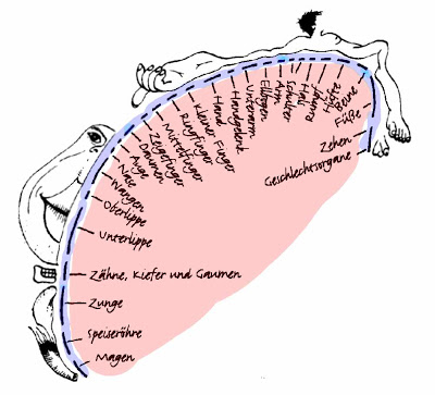
Homunculus image from Reinhard Blutner.
OK kids, let's start today's lesson by viewing the G-Rated [i.e., genital-less] flash explanation of homunculus.
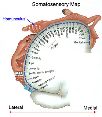
The neuroanatomical definition of homunculus is a "distorted" representation of the sensorimotor body map (and its respective parts) overlaid upon primary somatosensory and primary motor cortices. The above figure illustrates the sensory homunculus, where each body part is placed onto the region of cortex that represents it, and the size of the body part is proportional to its cortical representation (and sensitivity). It's rare to see the genitals represented at all. And if they are present, they are inevitably male genitals. To remedy this puritanical and androcentric situation, Swiss scientists at University Hospital in Zurich conducted a highly stimulating study in 15 healthy women to map the somatosensory representation of the clitoris (Michels et al., 2009).
The authors begin by reviewing the work of Wilder Penfield et al.:
The neuroimaging results revealed that compared to the rest blocks,

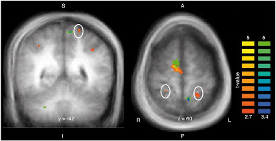
Fig. 3 (Michels et al., 2009). Illustration of the random-effect group-activation pattern for the contrast ‘electrical clitoral stimulation versus rest’ (orange–yellow color code; p less than 0.02 uncorrected for multiple comparisons) and for the contrast ‘electrical hallux stimulation versus rest’ (green–blue color code; p less than 0.001 uncorrected for multiple comparisons) on a group average brain. A cluster extent threshold of p less than 0.05 is applied for both contrasts. Electrical clitoral stimulation elicited bilateral activations of lateral surface of S1 as indicated by the white circles.
The major result was similar to the penile homuculus findings of Kell et al. (2005): a failure to replicate the original 1937 studies of Penfield and Boldrey. Although the statistical thresholds here for the clitoral stimulation were not stringent enough, the authors use this to their advantage:

Footnotes
1 Somatotopy, or somatotopic organization refers to
2 [like the standard homunculi shown above and below]
ReferencesKell CA, von Kriegstein K, Rösler A, Kleinschmidt A, Laufs H. (2005). The sensory cortical representation of the human penis: revisiting somatotopy in the male homunculus. J Neurosci. 25:5984-7.

Michels, L., Mehnert, U., Boy, S., Schurch, B., & Kollias, S. (2009). The somatosensory representation of the human clitoris: An fMRI study NeuroImage. DOI: 10.1016/j.neuroimage.2009.07.024
- Neurosurgeons Find Small Brain Region That Turns Consciousness On And Off, Like The Key In A Car's Ignition
The 54-year-old epilepsy patient - her name remains concealed to protect her privacy - was lying on the operating table while surgeons explored inside her brain with electrodes. They were looking for the source of her epileptic seizures. Suddenly, after...
- Targeted Brain Stimulation Provokes Feelings Of Bliss
It's hard to fathom how our subjective lives can be rooted in the spongy flesh of brain matter. Yet the reality of the brain-mind link was made stark half way through the last century by the Canadian neurosurgeon Wilder Penfield. Before conducting...
- The Purring Center In Cats
Large black spots show points from which stimulation elicited purring. Small black spots show points in these sections which were stimulated without eliciting purring. Numerous other points in other sections were stimulated with negative results so far...
- A New Penile Homunculus?
Homunculus image from Reinhard Blutner. Leave it to those wacky writers at LiveScience.com to come up with a sure-to-click headline: Study: Circumcision Removes Most Sensitive Parts By Ker Than, LiveScience Staff Writer posted: 15 June 2007 12:53 pm...
- Alternative To Deep-brain Stimulation In Parkinson Disease?
From Reuters:Brain Surface Stimulation May Ease Parkinson's Mon Jan 3, 2005 06:22 PM GMT By Anne Harding NEW YORK (Reuters Health) - Electrical stimulation of regions deep in the brain has become fairly common in recent years for treating Parkinson's...
Neuroscience
A New Clitoral Homunculus?

Homunculus image from Reinhard Blutner.
OK kids, let's start today's lesson by viewing the G-Rated [i.e., genital-less] flash explanation of homunculus.
The neuroanatomical definition of homunculus is a "distorted" representation of the sensorimotor body map (and its respective parts) overlaid upon primary somatosensory and primary motor cortices. The above figure illustrates the sensory homunculus, where each body part is placed onto the region of cortex that represents it, and the size of the body part is proportional to its cortical representation (and sensitivity). It's rare to see the genitals represented at all. And if they are present, they are inevitably male genitals. To remedy this puritanical and androcentric situation, Swiss scientists at University Hospital in Zurich conducted a highly stimulating study in 15 healthy women to map the somatosensory representation of the clitoris (Michels et al., 2009).
The authors begin by reviewing the work of Wilder Penfield et al.:
During the last 70 years the description of the sensory homunculus has been virtually a standard reference for various somatotopical studies (Penfield and Boldrey 1937; PDF). This map consists of a detailed description of the functional cortical representation of different body parts obtained via electrical stimulation during open brain surgery. In their findings they relied on reported sensations of different body parts after electrical stimulation of the cortex. Assessment of the exact location was generally difficult and sometimes led to conflicting results. The genital region was especially hard to assess due to difficulties with sense of shame.Recent studies have tried to map the somatosensory represenation of the human penis using neuroimaging methods, but there has been disagreement over whether it shows the classic medial representation seen in the figure above, or a more laterally located representation in the postcentral gyrus. For example, Kell et al. (2005) noted that...
...classical and [some] modern findings appear to be at odds with the principle of somatotopy,1 often assigning it to the cortex on the mesial wall. Using functional neuroimaging, we established a mediolateral sequence of somatosensory foot, penis, and lower abdominal wall representation on the contralateral postcentral gyrus in primary sensory cortex and a bilateral secondary somatosensory representation in the parietal operculum.But there are no comparable fMRI studies of female genitalia. So how is such a study conducted, methodologically speaking? Electrical stimulation of the dorsal clitoral nerve was compared to electrical stimulation of the hallux (big toe). It was all very clinical, no sexual arousal involved. Here's the experimental protocol:
Prior to the imaging session, two self-attaching surface disc electrodes (1 × 1 cm) were placed bilaterally next to the clitoris of the subjects so that we were able to stimulate the fibers of the dorsal clitoral nerve. Before the start of the experiment, electrical test stimulation was performed to ensure that subjects could feel the stimulation directly at the clitoris. In addition, the strength of electrical stimulation was adjusted to a subject-specific level, i.e. that stimulation was neither felt [as] painful nor elicited – in case of clitoris stimulation – any sexual arousal (see below). Functional imaging was performed in a block design with alternating rest and stimulation conditions, starting with a rest condition. ... In addition to the clitoris stimulation, we performed in eight of the recorded subjects a second experimental session, in which we applied electrical stimulation of the right hallux using the same type of electrodes, stimulation and scan paradigm.If you "see below" in the Methods you'll discover that after the fMRI session, participants rated their level of sexual arousal and discomfort on a visual analogue scale that ranged from -10 (unbearable pain or strong sexual arousal) to 10 (pleasure or no arousal at all/sleepiness). The median score for sexual arousal was zero with some variability [range: −7.5 to 8; −2 (25% percentile) and 2.5 (75% percentile)]. The median score for comfortableness was −2 [range: −7 to 9; −2.5 (25% percentile) and 0 (75% percentile)]. C'est la vie.
The neuroimaging results revealed that compared to the rest blocks,
Electrical clitoral stimulation produced significant activations predominantly in bilaterally prefrontal areas (BA 6, 8 and 45), the precentral, parietal and postcentral gyri, including S1 (BA 2 and 3; 40–70% probability) and S2 (BA 43 and ventral BA 40, 30–60% probability). In addition, distributed activations were also seen in the anterior and posterior parts of the insula and the putamen.

Fig. 3 (Michels et al., 2009). Illustration of the random-effect group-activation pattern for the contrast ‘electrical clitoral stimulation versus rest’ (orange–yellow color code; p less than 0.02 uncorrected for multiple comparisons) and for the contrast ‘electrical hallux stimulation versus rest’ (green–blue color code; p less than 0.001 uncorrected for multiple comparisons) on a group average brain. A cluster extent threshold of p less than 0.05 is applied for both contrasts. Electrical clitoral stimulation elicited bilateral activations of lateral surface of S1 as indicated by the white circles.
The major result was similar to the penile homuculus findings of Kell et al. (2005): a failure to replicate the original 1937 studies of Penfield and Boldrey. Although the statistical thresholds here for the clitoral stimulation were not stringent enough, the authors use this to their advantage:
We found no evidence of clitoral representation in the mesial wall, even when using unconventionally low statistical thresholds. This finding is further substantiated by other recent cytoarchitectonic studies revealing that BA 2 does not reach the inter-hemispheric fissure and BA 3 and BA 1 reach the postcentral mesial wall with a probability of only 30% . Our results are also in good agreement with [neuroanatomical] studies on nonhuman primates.In conclusion, it appears that Michels et al. (2009) have indeed mapped out a new clitoral homunculus, to go along with the new penile homunculus. The standard somatosensory images2 should be revised accordingly.

Footnotes
1 Somatotopy, or somatotopic organization refers to
the maintenance of spatial organisation within the central nervous system. For example, sensory information maintains its structure (i.e. sensory information on the hand remains next to sensory information on the arm) throughout the spinal cord and brain.Foot fetishes aside, the mapping of the genitals next to the toes is in violation of somatotopic organization.
2 [like the standard homunculi shown above and below]
ReferencesKell CA, von Kriegstein K, Rösler A, Kleinschmidt A, Laufs H. (2005). The sensory cortical representation of the human penis: revisiting somatotopy in the male homunculus. J Neurosci. 25:5984-7.

Michels, L., Mehnert, U., Boy, S., Schurch, B., & Kollias, S. (2009). The somatosensory representation of the human clitoris: An fMRI study NeuroImage. DOI: 10.1016/j.neuroimage.2009.07.024
- Neurosurgeons Find Small Brain Region That Turns Consciousness On And Off, Like The Key In A Car's Ignition
The 54-year-old epilepsy patient - her name remains concealed to protect her privacy - was lying on the operating table while surgeons explored inside her brain with electrodes. They were looking for the source of her epileptic seizures. Suddenly, after...
- Targeted Brain Stimulation Provokes Feelings Of Bliss
It's hard to fathom how our subjective lives can be rooted in the spongy flesh of brain matter. Yet the reality of the brain-mind link was made stark half way through the last century by the Canadian neurosurgeon Wilder Penfield. Before conducting...
- The Purring Center In Cats
Large black spots show points from which stimulation elicited purring. Small black spots show points in these sections which were stimulated without eliciting purring. Numerous other points in other sections were stimulated with negative results so far...
- A New Penile Homunculus?
Homunculus image from Reinhard Blutner. Leave it to those wacky writers at LiveScience.com to come up with a sure-to-click headline: Study: Circumcision Removes Most Sensitive Parts By Ker Than, LiveScience Staff Writer posted: 15 June 2007 12:53 pm...
- Alternative To Deep-brain Stimulation In Parkinson Disease?
From Reuters:Brain Surface Stimulation May Ease Parkinson's Mon Jan 3, 2005 06:22 PM GMT By Anne Harding NEW YORK (Reuters Health) - Electrical stimulation of regions deep in the brain has become fairly common in recent years for treating Parkinson's...
