Neuroscience

Is a laboratory test or brain scanning method for diagnosing psychiatric disorders right around the corner? How about a test to choose the best method of treatment? Many labs around the world are working to solve these problems, but we don't yet have such diagnostic procedures (despite what some might claim). A new study by McGrath et al. (2013) might be a step in that direction, but the results are very preliminary and await further validation.
The principal investigator of that study is Dr. Helen Mayberg, a leader in neuroimaging studies of major depression. She and her colleagues have pioneered the use of deep brain stimulation (DBS) as a treatment for severe, intractable depression, which was "the culmination of 15 years of research using brain imaging technology," says Dr. Mayberg.
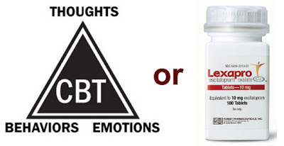
Psychotherapy or Drugs?
The choice of treatment modality in depression, as in other psychiatric disorders, is by trial and error. If one drug doesn't work, switch to another one. If your insurance covers it, a short course of evidence-based psychotherapy1 might be in order.
The whole concept of a DSM-based classification scheme for mental illnesses has come under fire, especially with the release of the new Diagnostic and Statistical Manual. In the real world, psychiatric disorders don't always show such clear boundaries; overlap and co-morbidity are common. The National Institute of Mental Health has endorsed a new approach, the Research Domain Criteria project, that incorporates dimensions of observable behavior along with neurobiological measures.
Here's where the new work by McGrath et al. (2013) fits in. Their goal was...
Fewer than 40% of depressed patients remit with their first course of treatment, so this would be an important advance. A more scientific way of choosing among possible treatment options would benefit patients and society at large.
The study (registered at clinicaltrials.gov, NCT00367341) enrolled a total of 82 depressed people. The neuroimaging method might surprise some of you: FDG-PET to measure glucose metabolism -- not the popular and trendy resting state fMRI to examine functional connectivity or any sort of fMRI activation study. However, the authors cite an established literature using this technique in studies of antidepressant treatment response.
Patients diagnosed with moderate to severe depression (a score of 18 or more on the Hamilton Depression Rating Scale, HDRS) received a PET scan and were randomized to receive 12 weeks of either cognitive behavioral therapy (CBT, n=41) or escitalopram (Lexapro, n=39), an SSRI antidepressant. Sixty-three patients completed this phase and also had a PET scan. The endpoint considered a successful response to treatment was remission (HDRS score of 7 or less), while non-response was a change in HDRS of 30% or less. Partial responders were omitted, leaving the final groups as follows:
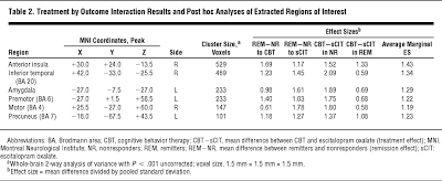
How was the biomarker identified? The PET images were co-registered with the corresponding structural MRIs. A whole brain analysis identified regions showing a treatment × outcome interaction (at a significance level of p<.001 uncorrected). Six regions met this uncorrected standard: right anterior insula, right inferior temporal cortex, left amygdala,2 left premotor cortex, right motor cortex, and precuneus (medial superior parietal lobe). Most of these are pretty surprising, but even more surprising is that the rostral anterior cingulate (and subgenual cingulate, BA 25) were not involved:

Effect sizes are shown in the table above. The brain regions were ranked in order of size of activation (which doesn't make sense for the amygdala), and the right anterior insula was chosen as the best potential biomarker because.... it had the largest cluster size? Or because it did marginally better than the other regions in terms of effect size (although this was not shown statistically). As a hub for interoceptive awareness, attention, and emotion, the anterior insula makes the most sense scientifically (Craig, 2009). Certainly, it would be odd if glucose metabolism in the right motor cortex could predict response to CBT or SSRI...
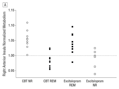
At any rate, right insula hypometabolism at baseline was associated with remission to CBT and poor response to SSRI, and vice versa for hypermetabolism. There was overlap between the groups as shown below, but increasing the chances of successful treatment (even with no guarantees) would be better than a completely trial-and-error approach.3
 Figure 3A (modified from McGrath et al., 2013). Right anterior insula as the optimal treatment-specific biomarker candidate. A. Scatterplot of insular activity from individual subjects in the remitter (REM) and nonresponder (NR) groups. Note: the anterior insula is the only region where the interaction subdivides patients into hypermetabolic (region/whole-brain mean >1.0) and hypometabolic (region/whole-brain mean <1.0) subgroups.
A Nature news story says that Brain scan predicts best therapy for depression, but that would be a premature conclusion at best. Although this study might be considered promising, the results must be validated in larger independent samples of patients who are assigned to treatments according to their baseline insula PET scans.
With the newly prominent nattering nabobs of neuroimaging negativity, it's important to remember that it's not all neuroprattle and bunk. Some of this research is trying to alleviate human suffering.
Further Reading
The Sad Cingulate
Sad Cingulate on 60 Minutes and in Rats
The Sad Cingulate Before CBT
Deep Brain Stimulation for Bipolar Depression
Is CBT Worthless?
Where Are the Clinical Tests for Psychiatric Disorders?
The Dark Side of Diagnosis by Brain Scan
ADDENDUM (6/14/2013): David Dobbs has an excellent post on the same study, Talk Therapy or Pill? A Brain Scan May Tell What’s Best. Dobbs has written extensively about Dr. Mayberg and her work, including A Depression Switch? – New York Times and Depression’s wiring diagram.
Footnotes
1 But read LawsDystopiaBlog by Professor Keith Laws to see how flimsy the "evidence base" can sometimes be.
2 An earlier experiment showed that the amygdala might be a region that could help predict CBT response, using fMRI and response to emotional words.
3 Not to be a pedantic stick in the mud, but the combination of drugs and therapy is often the most successful.
References
Craig AD. (2009). How do you feel--now? The anterior insula and human awareness. Nat Rev Neurosci. 10:59-70.
McGrath CL, Kelley ME, Holtzheimer PE, Dunlop BW, Craighead WE, Franco AR, Craddock RC, & Mayberg HS (2013). Toward a Neuroimaging Treatment Selection Biomarker for Major Depressive Disorder. JAMA psychiatry (Chicago, Ill.), 1-9 PMID: 23760393
- Broaden Trial Of Dbs For Treatment-resistant Depression Halted By The Fda
Webpage for the BROADEN™ study formerly run by St. Jude Medical It's become mainstream these days to say that psychiatric disorders are neural circuit disorders. You can even read all about it in the New York Times! Cognitive training and neuromodulation...
- Good News/bad News Update On Nucleus Accumbens Dbs For Treatment-resistant Depression
Taken from Fig. 1 (Bewernick et al., 2009). Hamilton Depression Rating Scale (PDF) over time. Two and a half years ago, The Neurocritic wrote about the very early results of deep brain stimulation (DBS) in the nucleus accumbens for severe, refractory...
- ...but My Subgenual Cingulate Is Sad
In the previous post, The Neurocritic discussed an article in Nature in which greater activity in the rostral anterior cingulate cortex (rACC) was associated with optimism1 (Sharot et al., 2007). "But wait!" you might say, "what about the sad cingulate?"...
- Sad Cingulate On 60 Minutes And In Rats
Happy to have stereotactic brain surgery while conscious! Today is National Depression Screening Day, and the sad cingulate (ventral anterior cingulate cortex, or Brodmann area 25) is in the news again. 60 Minutes did a piece on two patients undergoing...
- The Sad Cingulate Before Cbt
The latest sad cingulate news is an fMRI study that examined the responsiveness of this region (subgenual cingulate cortex, aka Brodmann area 25) to emotional stimuli as a predictor of recovery in depressed patients receiving cognitive behavior therapy...
Neuroscience
A New Biomarker for Treatment Response in Major Depression? Not Yet.

Is a laboratory test or brain scanning method for diagnosing psychiatric disorders right around the corner? How about a test to choose the best method of treatment? Many labs around the world are working to solve these problems, but we don't yet have such diagnostic procedures (despite what some might claim). A new study by McGrath et al. (2013) might be a step in that direction, but the results are very preliminary and await further validation.
The principal investigator of that study is Dr. Helen Mayberg, a leader in neuroimaging studies of major depression. She and her colleagues have pioneered the use of deep brain stimulation (DBS) as a treatment for severe, intractable depression, which was "the culmination of 15 years of research using brain imaging technology," says Dr. Mayberg.
Psychotherapy or Drugs?
The choice of treatment modality in depression, as in other psychiatric disorders, is by trial and error. If one drug doesn't work, switch to another one. If your insurance covers it, a short course of evidence-based psychotherapy1 might be in order.
The whole concept of a DSM-based classification scheme for mental illnesses has come under fire, especially with the release of the new Diagnostic and Statistical Manual. In the real world, psychiatric disorders don't always show such clear boundaries; overlap and co-morbidity are common. The National Institute of Mental Health has endorsed a new approach, the Research Domain Criteria project, that incorporates dimensions of observable behavior along with neurobiological measures.
Here's where the new work by McGrath et al. (2013) fits in. Their goal was...
To identify a candidate neuroimaging “treatment-specific biomarker” that predicts differential outcome to either medication or psychotherapy.
Fewer than 40% of depressed patients remit with their first course of treatment, so this would be an important advance. A more scientific way of choosing among possible treatment options would benefit patients and society at large.
The study (registered at clinicaltrials.gov, NCT00367341) enrolled a total of 82 depressed people. The neuroimaging method might surprise some of you: FDG-PET to measure glucose metabolism -- not the popular and trendy resting state fMRI to examine functional connectivity or any sort of fMRI activation study. However, the authors cite an established literature using this technique in studies of antidepressant treatment response.
Patients diagnosed with moderate to severe depression (a score of 18 or more on the Hamilton Depression Rating Scale, HDRS) received a PET scan and were randomized to receive 12 weeks of either cognitive behavioral therapy (CBT, n=41) or escitalopram (Lexapro, n=39), an SSRI antidepressant. Sixty-three patients completed this phase and also had a PET scan. The endpoint considered a successful response to treatment was remission (HDRS score of 7 or less), while non-response was a change in HDRS of 30% or less. Partial responders were omitted, leaving the final groups as follows:
- CBT remission, n=12
- escitalopram remission, n=11
- CBT nonresponse, n=9
- escitalopram nonresponse, n=6
How was the biomarker identified? The PET images were co-registered with the corresponding structural MRIs. A whole brain analysis identified regions showing a treatment × outcome interaction (at a significance level of p<.001 uncorrected). Six regions met this uncorrected standard: right anterior insula, right inferior temporal cortex, left amygdala,2 left premotor cortex, right motor cortex, and precuneus (medial superior parietal lobe). Most of these are pretty surprising, but even more surprising is that the rostral anterior cingulate (and subgenual cingulate, BA 25) were not involved:
Contrary to past published studies,63 the rostral anterior cingulate did not discriminate the outcome subgroups in either the main effect or interaction analyses. A post hoc examination of responder and nonresponder differences within each treatment arm did reveal a nonsignificant rostral cingulate activity difference, with metabolism in responders greater than nonresponders, but solely in the escitalopram group. While consistent with past reports, this finding did not meet the TSB [treatment-specific biomarker] criteria defined for the current study, ie, a region whose activity can differentiate both good and poor outcomes for both treatments.
- click on image for a larger view -

Effect sizes are shown in the table above. The brain regions were ranked in order of size of activation (which doesn't make sense for the amygdala), and the right anterior insula was chosen as the best potential biomarker because.... it had the largest cluster size? Or because it did marginally better than the other regions in terms of effect size (although this was not shown statistically). As a hub for interoceptive awareness, attention, and emotion, the anterior insula makes the most sense scientifically (Craig, 2009). Certainly, it would be odd if glucose metabolism in the right motor cortex could predict response to CBT or SSRI...
At any rate, right insula hypometabolism at baseline was associated with remission to CBT and poor response to SSRI, and vice versa for hypermetabolism. There was overlap between the groups as shown below, but increasing the chances of successful treatment (even with no guarantees) would be better than a completely trial-and-error approach.3

A Nature news story says that Brain scan predicts best therapy for depression, but that would be a premature conclusion at best. Although this study might be considered promising, the results must be validated in larger independent samples of patients who are assigned to treatments according to their baseline insula PET scans.
With the newly prominent nattering nabobs of neuroimaging negativity, it's important to remember that it's not all neuroprattle and bunk. Some of this research is trying to alleviate human suffering.
Further Reading
The Sad Cingulate
Sad Cingulate on 60 Minutes and in Rats
The Sad Cingulate Before CBT
Deep Brain Stimulation for Bipolar Depression
Is CBT Worthless?
Where Are the Clinical Tests for Psychiatric Disorders?
The Dark Side of Diagnosis by Brain Scan
ADDENDUM (6/14/2013): David Dobbs has an excellent post on the same study, Talk Therapy or Pill? A Brain Scan May Tell What’s Best. Dobbs has written extensively about Dr. Mayberg and her work, including A Depression Switch? – New York Times and Depression’s wiring diagram.
Footnotes
1 But read LawsDystopiaBlog by Professor Keith Laws to see how flimsy the "evidence base" can sometimes be.
2 An earlier experiment showed that the amygdala might be a region that could help predict CBT response, using fMRI and response to emotional words.
3 Not to be a pedantic stick in the mud, but the combination of drugs and therapy is often the most successful.
References
Craig AD. (2009). How do you feel--now? The anterior insula and human awareness. Nat Rev Neurosci. 10:59-70.
McGrath CL, Kelley ME, Holtzheimer PE, Dunlop BW, Craighead WE, Franco AR, Craddock RC, & Mayberg HS (2013). Toward a Neuroimaging Treatment Selection Biomarker for Major Depressive Disorder. JAMA psychiatry (Chicago, Ill.), 1-9 PMID: 23760393
- Broaden Trial Of Dbs For Treatment-resistant Depression Halted By The Fda
Webpage for the BROADEN™ study formerly run by St. Jude Medical It's become mainstream these days to say that psychiatric disorders are neural circuit disorders. You can even read all about it in the New York Times! Cognitive training and neuromodulation...
- Good News/bad News Update On Nucleus Accumbens Dbs For Treatment-resistant Depression
Taken from Fig. 1 (Bewernick et al., 2009). Hamilton Depression Rating Scale (PDF) over time. Two and a half years ago, The Neurocritic wrote about the very early results of deep brain stimulation (DBS) in the nucleus accumbens for severe, refractory...
- ...but My Subgenual Cingulate Is Sad
In the previous post, The Neurocritic discussed an article in Nature in which greater activity in the rostral anterior cingulate cortex (rACC) was associated with optimism1 (Sharot et al., 2007). "But wait!" you might say, "what about the sad cingulate?"...
- Sad Cingulate On 60 Minutes And In Rats
Happy to have stereotactic brain surgery while conscious! Today is National Depression Screening Day, and the sad cingulate (ventral anterior cingulate cortex, or Brodmann area 25) is in the news again. 60 Minutes did a piece on two patients undergoing...
- The Sad Cingulate Before Cbt
The latest sad cingulate news is an fMRI study that examined the responsiveness of this region (subgenual cingulate cortex, aka Brodmann area 25) to emotional stimuli as a predictor of recovery in depressed patients receiving cognitive behavior therapy...
